Sunday, April 21, 2024
Shouder Keeps Clicking But No Pain
Sunday, November 27, 2022
Will Marathon Running Hurt Your Back?
| Spot my compression fracture at L1? |
| My L1 is 'collapsed' compared to above and below |
Sunday, January 30, 2022
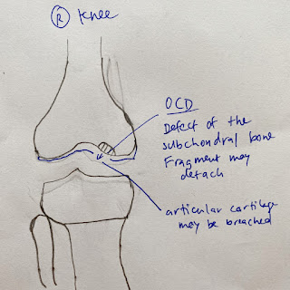
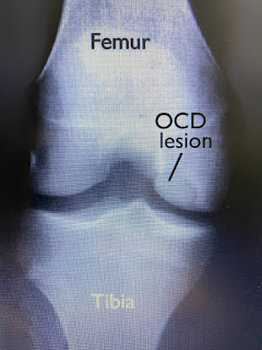
OCD
| Kindly drawn by my older son |
The cause of OCD is unknown. It is suggested that reduced blood flow to the bone may occur from repetitive trauma like excessive running and jumping. Or small, multiple episodes of minor unknown injury that damage the bone over time. Some studies suggest that there may be a genetic component involved too.
OCD occurs most frequently in children and adolescents, most commonly in the knee, but can also occur in the elbow and ankle.
Doctors stage OCD according to how big the bone fragment is, whether the bone fragment is totally or partially separated or whether it stays in place. If that loosened piece of bone is not detached, then there may be few or no symptoms. For younger children whose bones are still growing and developing, there is a chance that it can heal.
So it was a total surprise to me when my adult patient was diagnosed with OCD 2 days ago.
She was in the process of sitting down on her sofa and slightly twisted her knee. She could not extend or bend her knee fully nor put weight on her foot after that. She went to the hospital and was told it might be a meniscus tear and may need surgery. The surgeon suggested an MRI to confirm his hypothesis.
Hence, when the MRI results came out it was a big surprise that there was a bone fragment dislodged. That's the reason why she could not straighten or bend her knee fully.
The only incident my patient could think of was during gymnastics, when she was 12, that her knee started to hurt (possibly from the impact of repeated running and landing). Her knee would 'lock' occasionally for a few days at a time. It gradually resolved and she would occasionally have some knee pain on that same left knee while she competed in triathlons. Otherwise, there was no other clue that she would have OCD.
Now my patient has to decide how she would like to proceed to get the bone fragment out after discussion with her surgeon.
Reference
Kocher MS, Tucker R, Ganley TJ et al (2006). Management Of Osteochondritis Dissecans Of The Knee: Current Concepts Review. AJSM. 34(7): 1181-1191. DOI: 10.177/0363546506290127.
![]() |
| Picture from RadioGraphics |
Sunday, January 2, 2022
Pain Does Not Mean You're Injured
Most elite athletes I treat are similar, always dealing with pain and niggles. Some pain would disappear while other pain tend to persist and linger.
A recent article (Hoegh et al, 2021) suggests that in the context of sports medicine, pain and injuries are 2 different distinct entities and should not be lumped together.
That article (Hoegh et al, 2021) suggests that when pain is inappropriately labeled as an injury, it creates fear and anxiety. It may even change how we move the affected body part, creating further problems.
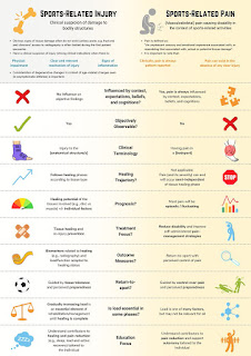
| From Hoegh et al, 2021 |
Reading words like stabbing pain or burning sensations can affect how you feel. When we complain of pain, it may feel like something is damaged. However, as I often tell my patients, pain is subjective. To my patient it may feel like a 3 out of 10 kind of pain, but to me it may be an 8 out of 10 pain. Or vice versa. Pain can occur and exist even without an injury.
Pain can be influenced by beliefs, the process of cognition (knowing and perceiving), expectations and circumstances. Injuries are not. The onset of pain can be unpredictable, and how severe the pain is does not usually depend on the stage of healing.
Injuries can be identified by sight, orthopaedic tests and scans. The prognosis for an injury will depend on where the injury is. A muscle tear will usually heal faster than a bone fracture.
After a sports injury, in order for the athlete to return to sport, we gradually increase their training load on their damaged tissue during rehabilitation until healing is complete and able to handle the demands of more strenuous training and competition.
With sports related pain, one cannot gradually increase training load and hope that the pain will go away. However, we can improve the patient's ability to manage the pain especially since pain is subjective.
If you have a bone stress fracture, you will have to take time off running until it heals and gradually increase load. Once healed, pain usually is no longer an issue. The injury and the associated pain are tightly connected.
Other cases may not be as straight forward. An example would be patients who have chronic pain in their Achilles tendon with no damage on their MRI scans. There is no clear link between the physical state of their tendon and how it feels (or hurts). In such cases, surely we can manage and reduce their pain to allow them to run rather than waiting for their tendon to 'heal'.
Another recent publication by Friedman et al (2021) warn of the dangers of diagnostic labels. Calling a patient's knee injury a meniscal tear rather than a meniscal strain may nudge the patient to opt for arthroscopic surgery even though that may not be considered the best approach.
According to the authors (Friedman et al, 2021), words chosen by medical professionals to describe injuries may present a situation as considerably worse than it actually is. This will increase anxiety and cause fear of movement.
Judging what pain to ignore and which ones to take seriously can be a delicate art rather than science and how we choose to label it can affect the outcome.
Sometimes pain is just pain.
References
Friedman DJ, Tulloch D, and Khan KM (2021). peeling Off Musculoskeletal Labels: Sticka and Stones May Break My Bones, But Diagnostic Labels Can Hamstring Me Forever. BJSM. 55: 1184-1185. DOI: 10.1136/bjsports-2021-103998.
Hoegh M, Stanton T, George S et al (2021). Pain Or Injury? Why Differentiation Matters In Exercise And Sports Medicine. BJSM. DOI: 10.1136/bjsports-2021-104633.
Sunday, October 4, 2020
What Happens When Patients Are Sent Too Early For An MRI
![]() |
| Picture by Naiserie from Flickr |
I was surprised, after examining him, that he was referred for an MRI so soon. His back pain did not seem sinister. At the end of yesterday's session, his back was completely pain free.
I shared with him a really interesting article I had just come across. The researchers studied data of patients seeking treatment for non-specific low back pain without a red flag (warning or danger) condition and no low back pain in the previous six months.
More patients had back surgery if they were referred for an MRI within the first six weeks of an initial visit to the doctor (1.48 % versus 0.12 % in cases without an early MRI).
The patients also complained of a higher pain score when they had an early MRI. In fact, overall outcomes were worse, including greater use and potential harm for prescription medication (35.1 % versus 28.6 %). There were also higher costs for other medical care ($8,802 versus $5,560).
This association was also true when patients had to pay for their treatment (compared with not having to pay at all).
Perhaps this information will help bring down costs for Singapore's Integrated Shield Plans since there was such an outcry when it was announced that premiums were going higher despite increasing coverage.
Reference
Jacobs JC, Jarvik JG et al (2020). Observational Study Of The Downstream Consequences Of Inappropriate MRI Of The Lumbar Spine. J Gen Int Med. DOI: 10.1007/s11606-020-06181-7.
Monday, February 10, 2020
Slipped Discs Can Heal
![]() |
| Disc strongly attached to vertebral end plate |
Since the IVD is part of our human anatomy just like our muscles, nerves, bones, tendons, ligaments etc, the IVD can heal with correct conservative management and time. Consider the following evidence.
Chui et al (2015) did a systematic review of the literature to look for evidence of our lumbar discs healing. Their inclusion criteria was patients who had herniated lumbar discs that were treated conservatively and patients must have at least two imaging evaluations of their lumbar spine.
Patients who had previous lower back surgery, tumours, spinal infections, spondylolistheis and spinal stenosis were excluded from the review. In their systematic review, the authors found 31 studies that fit their criteria.
Their results will definitely surprise you. Here's what the author's wrote. "Spontaneous regression of herniated disc tissue can occur, and can completely resolve after conservative treatment. Patients with disc extrusion and sequestration had a significantly higher possibility of having spontaneous regression than did those with bulging or protruding discs. Disc sequestration had a significantly higher rate of complete regression than dis disc extrusion."
The larger the disc injury, the more likely it is to revert to its former shape and heal! For disc sequestration, rate of spontaneous regression was found to be 96%. For disc extrusion 70%, 41% for disc protrusion and 13% for a disc bulge.
It does seem like (my personal opinion) the more serious the disc injury, the larger response our bodies have for healing.
In another study by Nakashima et al (2015), the authors studied the MRI's of 1211 asymptomatic adults ranging from 20 to 70 years old. They found that 87.6% of subjects presented with disc bulges, and this significantly increased with age in terms of frequency.
Even most of the subjects in the 20's had bulging discs, 73.3% of males and 78% of females respectively. The authors concluded that disc bulging was frequently observed in asymptomatic subjects including those in their 20's.
It is my hope that after reading this, patients who have been told that they have on their MRI 'slip' discs, herniated discs etc will have new found knowledge and confidence that they can definitely get better with conservative management and not require surgery.
If the healthcare professional treating your 'slip disc' tells you you need surgery, you can show them this article.
References
Chui CC, Chuang TY et al (2015). The Probability Of Spontaneous Regression Of Lumbar Herniated Disc: A Systematic Review. Clin Rehabil. 29(2); 184-195. DOI: 10.1177/0269215514540919
Nakashima H, Yukawa Y et al (2015). Abnormal Findings On Magentic Resonance Images Of The Cervical Spines In 1211 Asymptomatic Subjects. Spine. 40(6): 392-398. DOI: 10.1097/BRS.0000000000000775
Sunday, January 5, 2020
Excuse Me, Are You A Physiotherapist?
![]() |
| At Pippa's b'day this morning |
I used to, but not anymore. Every time I tell somebody I meet I'm a physiotherapist they end up asking me about a pain or condition they have. Even friends that I haven't met for a while will ask me about their pain before they've even said hello sometimes.
Don't get me wrong, I'm not complaining. And it's not because I don't want to help or prefer them to come to our clinic so I can charge them. It's natural to ask someone who knows what they're doing for an opinion. Hey, I do the same pretty much all the time.
I often ask my patients who work in financial markets whether the stock market is heading up or down. Or if the US dollar is appreciating. Or is it time to buy some gold or silver bullion?
![]() |
| Picture from USAGold |
He added that stock tip hunters never consider what time horizon lies behind the idea/ tip as they always assume the information leads to a "quick buck".
As a physiotherapist/ health care professional, I definitely do not mind helping someone who asks look for a pathway to solve their problem. For instance, which specialist to consider, which hospital that has the best facilities, MRI scan especially if the problem is beyond me.
![]() |
| Comic from Pinterest.com |
For a clinical diagnosis is a well-informed weighing of probabilities. No diagnosis has a 100 percent certainty unless you can assess the patient physically.
If you do happen to ask for my thoughts on your pain/ condition (at a birthday party etc), I'll still give my opinion but will definitely suggest a physical assessment to follow up in our clinic or get another health professional to verify.
Happy New Year.
Another funny comic from Pinterest
Sunday, December 22, 2019
$677 For A Session Of Acupuncture Or Physiotherapy!!
![]() |
| Posed picture |
I had to politely decline as I thought it wasn't right to make insurance pay for the scan if a patient does not want to foot the bill.
So I'm not surprised to read that there are mercenary physiotherapists, chiropractors etc who take advantage of the insurance system to bill excessively.
In New Jersey, school employees are covered by the state's School Employees' Health Benefits Program. This plan covering school teachers covers virtually anything the "out-of-network" providers charge.
In 2018, the teachers' plan paid out an average of more than USD $600 per visit to an acupuncturist or physiotherapist! This is even higher than out-of-network fees of psychiatrists and gynecologists.
As a result, more than 70 acupuncturists and physiotherapists earned more than USD $200,000 in 2018 from their teacher clients alone. Data also showed that one acupuncturist earned more than USD $1 million dollars!!
To further sweeten the deal, some of these providers are suspected of waiving a co payment by the teachers so there is no out of own pocket payment by the teachers.
The same state fund paid out USD $11.2 million dollars to a clinic -Thompson Healthcare and Sports Medicine for providing chiropractic services, acupuncture and physiotherapy to teachers.
![]() |
| Thompson Healthcare and Sports Med during a school visit |
Six people (half are union members) make up the state panel overseeing the benefit plan have not done anything to stop the runaway costs. This is despite meeting transcripts showing discussions regarding this since 2014.
All these data and documents, along with interviews with state officials show this is what happens when savvy (and mercenary) health care practitioners find lucrative loopholes in a loosely designed employee health plan.
As expected, the teachers' premiums have increased by 8 and 13 percent in the last two years. The most popular family premium plan cost in excess of $36,000 a year. This is nearly twice the cost in other parts of the country.
I dare say that if the teachers realized the exploitation of the plan, they will definitely think twice about how they use their health care benefits.
Back in in Singapore, there was an article in the Today online paper regarding some physiotherapists in private practices paying doctors and surgeons kickbacks to get patients referred to them.
You can also read our other blog, where we have previously written about these kickbacks in our Singapore healthcare system too. Needless to say, private medical clinics and MRI / X-ray companies in Singapore are implicated as well.
It is bad practices like this that will lead to increases in our health care visits, insurance premiums and Medisave-approved Integrated shield plans.
You can read the read I referenced here.
Monday, November 25, 2019
The Correct Touch Is Really Important
My patient said to me that she will never see that doctor again. She was annoyed that despite her complaining of pain in her knees, he did not even bother to examine her. There was no eye contact from him at all too. He told her brusquely that she had runner's knee and should not be running. The Sports Doctor then prescribed her some pain medication and referred her for physiotherapy.
She had gone to the physiotherapist referred but did not get better. Her friend suggested that she come to our clinic since the referred physiotherapist was not a runner as well. So here she was in our clinic.
Like I've written previously, I can never be totally sure of a patient's condition if I don't assess them properly. All patients need and deserve gentle and thoughtful treatment. However, healthcare is evolving and become more businesslike. However, that human touch is still really important.
We should never take for granted that our patients give us consent to touch them to be able to assess them. Especially now in an era of electronic medical records, doctors and other health care practitioners may spend most of the allocated appointment time staring at a computer screen, tablet or looking at MRI reports.
I'm so glad that Dr Abraham Verghese, a professor at Stanford University's School of Medicine still prefers "the bedside chat, the old fashioned physical exam and the power of informed observation".
In his 2011 TED Talk, he said that by shortening the physical examination, doctors will lose a ritual that is transformative, transcendent and at the heart of the physician patient relationship.
Of course there are instances when patients are in pain and/or anxious, they will be much more guarded. Definitely patients of sexual abuse or assault may cringe at the lightest touch.
I have on many different occasions seen a patient come in for say neck pain but had a worried look that seems disconnected from their physical problem. After assessing and/or treating them and asking them if they are alright they become tearful and mention a family death, possible relationship problems or financial difficulties. That placing of hands, that human touch was the catalyst that allowed the patient to open up and release the pent up emotions.
With that human touch, we can comfort, diagnose and bring about treatment.
Be careful with your touch. It can show gentleness, compassion and heal someone. Or it can show roughness, carelessness or even incompetence.
Sunday, June 2, 2019
Change The Arms, Change The Neck
![]() |
| First assessment |
This patient works as an electrician and often has to be in awkward positions running electrical wires. He feels worse after prolonged time spent looking upwards - mostly due to running ceiling electrical wires.
He was referred to our clinic by another friend who had seen us and gotten better without needing surgery as suggesting by his surgeon.
Just like the other lady who had neck pain, I didn't treat his neck. Just treated his arms and shoulder girdle.
Have a look at the picture after treatment.
![]() |
| After treatment |
![]() |
| Before and after |
What did I do? No mobilizations or manipulations of the thoracic and cervical spine at all. Just treating the arm lines as seen in the picture below except for levator scapulae. Left that out as I wanted to "exclude" the neck.
![]() |
| Superficial and deep back, front arm lines |
Friday, May 17, 2019
Not The Neck, Not The Leg, But The Arms
This patient who came in recently this week complaining of neck pain, was a really interesting case.
She had pain in the cervical spine (neck) area with numbness and tingling sensations radiating down to her right arm and thumb. Sleeping on her right side made the pain worse.
Remember I had written about my patient with neck pain and I made her better by treating her leg. Well, this particular patient's presentation was definitely different.
This patient of mine lives on a yacht in the One 15 Marina Sentosa Cove. Recently it's been raining and she's had to work a lot harder cleaning her yacht so there won't be stains. Because of the proximity of the refinery nearby, whenever it rains it makes their yacht very dirty. Her husband cleans the hull, and she needs to polish the silicone joints.
She had also seen the spine surgeon and he sent her for an MRI just to be sure. Here's the whatsapp message I received from the doctor telling me my patient had R spinal exit stenosis from prolapsed discs in her neck.
That was a big clue for me as her neck didn't didn't seem particularly "bad" upon palpation. Her upper limb tension tests elicited some tenderness as well.
![]() |
| Superficial and deep front, back arm lines |
When she sat up, her first comment was "it's like a whole weight have been lifted off my shoulders".
Surprised? I was, but I was hoping for a good result since she told me about all the extra cleaning she did earlier. Treating her arms was key in her case, not her neck.
Friday, August 10, 2018
Fat Pad Most Painful In The Knee?
His MRI results was like in his words "opening a can of worms" telling him what's wrong with his knees and perhaps that's why he started having pain after that.
After his ranting, I had to explain very thoroughly about the structures in our knees that cause the most pain. The information I gave him was derived from an article published quite a while ago in the American Journal of Sports Medicine but still very relevant today.
The doctors in that study came up with a simple method to document the various sensations felt inside a single subject's knees one week apart. Right knee first, followed by the left a week later. (Note that the subject had no prior knee pain).
They would arthroscopically poke/ palpate (using a specially built spring loaded device) different structures inside the knee while video recording the procedure and record what the subject's response was. Force used was between 0 to 500 grams. All this done without intra articular anesthesia. Ouch! That must really hurt.
The doctors only injected local anesthesia at the portal site (incision). The first author inspected both knees arthroscopically. He asked the patient when he poked at different structures and graded the sensation as follows (0) no sensation; (1) was non painful awareness; (2) slight discomfort; (3) moderate discomfort and (4) severe pain. This was done with with a modifier of either accurate spatial localization (A) or poor spatial localization (B).
Ready for the results? They were exactly the same for both knees. Even though it was done one week apart.
Palpation of the patellar articular cartilage in the three under surfaces (central ridge, medial and lateral facets) resulted in no sensation, or a 0 score, even with a strongest force of 500 grams. Palpation of the odd facets elicited a score of 1B. Asymptomatic grade II or III chondromalacia (wearing out) of the central ridge was identified on both patellas of the subject!
Palpation of the articular cartilage surfaces of the femoral condyles, trochlea, and tibial plateaus at 500 g of force universally produced a sensation of 1B to 2B.
The sensation from the meniscus ranged from 1B on the inner rim of the meniscus to to 3B near the capsular margin.
Sensation from the cruciate liagaments (Anterior, posterior cruciate ligaments) range from 1-2B in the mid-portion of the ligaments and 3-4B at the insertion sites.
Palpation of the suprapatellar pouch, capsule, and the medial and lateral retinacula produced a score of 3A to 4A (moderate to severe localized pain) at relatively low levels of force (about 100 g).
The most painful structures were the anterior synovium of the knee, the fat pad and the joint capsule - 4A.
The human knee can be very complex, especially our patellofemoral joint (patella and the femur). The three asymnetrical surfaces on the underside of the patella (or knee cap) has to work together with the femur as it accepts, transfers and dissipates loads between the bones.
We know from previous research that various structures in the knee send neurosensory signals (or messages) to the brain. It is theses signals that result in us feeling pain.
Even though my patient's patella cartilage had worn out (just like the subject) there shouldn't be any pain there as articular cartilage doesn't have any nerve supply. No nerve endings means it is unable to detect pain.
Even the ACL and meniscus wasn't really that sensitive to the poking. This observation may provide an explanation for the often poor localization of structural damage that many patients experience with a cruciate ligament or meniscal injury.
Now you know, worn out articular cartilage doesn't cause you pain. The pain you have is likely to come from other structures. And you definitely don't need to ingest any glucosamine too.
Reference
SF Dye, GL Vaupel and CC Dye (1998). Conscious Neurosensory Mapping Of The Internal Structures Of The Human Knee Without Intraarticular Anesthesia. AM J Sp Med. 26(6): 773-777. DOI: 10.1177/03635465980260060601.
![]() |
| black and white version |
Friday, June 30, 2017
How Is Running Good For Your Back?
Many of us runners have heard that running is bad for your knees (although I've put that beyond reasonable doubt here).
Many more of us have been told that running "overloads" our intervertebral discs (IVD) and causes jarring on our spine. And that in turn leads to low back pain.
I've written about how my own back feels better after running after my accident.
Well, here's more proof that people who regularly run or walk briskly tend to have healthier discs in their spines than people who do not exercise.
This findings refute the myth that running overloads your spine. In fact it shows that running makes the spine sturdier.
The IVD's are located between the vertebrae, acting as cushions to dissipate shock. They contain a thick, sticky fluid that compresses and absorbs pressure during movement to keep your spine in good shape.
Aging, disease and/ or injury can cause the IVD's to degenerate and bulge causing back pain which sometimes can be debilitating.
Check out the evidence provided in the following study. 79 adult men and women were recruited for the study, of which two-thirds of the group were runners for at least five years. The "long distance" group ran more than 30 miles (48 km) a week while the others ran between 12-25 miles a week (19-40 km). The last group rarely exercised at all.
In order to get more information out of the study, the subjects wore accelerometers. Accelerometers measure movement in terms of acceleration forces, or how much power your body is generating when you move.
All the subjects' spines were scanned using MRI, measuring size and liquidity of each disc. In general, the runners' discs were larger and contained more fluid than those who didn't exercise!
Mileage did not matter. The IVD's of the runners who ran less than 30 miles per week were almost identical to the "long distance" group. The authors suggested that compared to moderate mileage, heavy training does not increase disc health nor does it contribute to deterioration.
Here's what's more surprising. The accelerometers showed that walking briskly at about four miles (or 6.4 km) per hour generated enough physical force to bring movement into the range associated with the healthiest IVD's.
Slower walks and standing in place were outside this range. (Now you know why your backs hurt when you stand and not move). Running faster than 5.5 miles (or 8.8 km) per hour were outside the range as well.
The "sweet spot" for IVD's health seem to be between fast walks and gentle jogs.
Things to note. This is a one-time snapshot of the subject's backs. This study cannot prove that running (or exercise) caused the subjects' IVD's to become healthier. Not yet anyway. It shows that people who ran had healthier IVD's.
It also does not tell us whether running (or exercise) can help treat existing disc problems.
My thoughts? The available evidence strongly indicates that IVD's like movement. If you've always been walking and running don't listen to the naysayers. If you have never ran before and want to, perhaps it will help if you start walking briskly first, this will strengthen your IVD's. Progress to run walks (run a little, walk a little) before running to gradually ease your back into it.
Reference
Belavy DL, Quittner MJ, Ridgers N et al (2017). Running Exercise Strengthens The Intervertebral Disc. Scientific Reports. Article No: 45975. DOI: 10.1038/sreo45975.
Sunday, July 31, 2016
Do You Really Need An MRI?
![]() |
| Picture by Cory Doctorow from Flickr |
Results show she had a prolapsed disc (PID) or what is commonly as a slipped disc. The surgeon suggested surgical intervention to remove the disc but my patient refused and sought a second opinion.
Subsequently she came to our clinic after her friend with similar findings on MRI (but didn't have surgery) got better after seeing me.
I've encountered some patients who showed up in our clinics with "many problems" on their MRI but no pain while other patients showed no abnormality on their MRI but complain of severe pain. If you "believe" the MRI you may end up treating the MRI and not treating the patient (or trying to fix the problem on the MRI rather than addressing the actual issue causing your patients' symptoms/ pain).
Personally, I believe that nine times out of ten, a competent health care provider (doctor, physiotherapist etc) can pinpoint the cause of your pain/ injury without ordering an expensive MRI.
In our clinics, we ask many questions about the patients' symptoms, training regime (for a sports injury), much like a detective looking for clues. We then do a thorough physical assessment - comparing limbs, palpating the area that hurts, moving your limbs/ joints through different positions or have the patient perform the aggravating movement, checking alignment etc.
Done correctly and accurately, we can often pinpoint the root cause of the problem from the physical assessment and treat it.
It's also interesting to note that everyone is built a little differently and our structures change with age. As MRI's are very sensitive, they can reveal abnormalities that aren't the actual cause of your problems.
A recent review article found 37 percent of 20 year old subjects and 96 percent of 80 year old subjects have evidence of disc degeneration on MRI. The authors concluded imaging findings of spine degeneration are present in high proportions of individuals with no pain. These changes in the spine may be a sign of normal ageing rather than medical conditions/ acute injuries that require treatment.
Here's a common running related example. Another of my patients came to our clinic after seeing a Traditional Chinese Physician, physiotherapist and even saw a surgeon for medial (inside) heel pain and didn't get better. She had been walking more than normal and woke up having to hobble with pain upon setting her foot on the ground. That clearly to me would lead me to check her plantar fascia.
In fact, her surgeon did order an MRI and confirmed what I suspected. He proceeded to give her a cortisone injection (steroid injection) which didn't help. My patient endured the cost and hassle of doing the scan for no good reason.
I treated her twice and am happy to report she's well on her way to recovery.
Don't get me wrong. There are definitely times when a MRI scan is needed. If you've had a Physiotherapist /doctor etc assess you thoroughly, rested, had treatment and still not gotten better then it might be a good time to get an ultrasound scan, x-ray or MRI to investigate further.
What about the first patient who had a PID whom I wrote about at the start of this post? Well, she's back running, weight training like normal. Her doctor was surprised to say the least ......
Reference
Brinjkii W, Luetmer PH et al (2015). Systematic Literature Review Of Imaging Features Of Spinal Degeneration In Asymptomatic Populations. J Am Neuroradiol. 36(4): 811-816. DOI: 10.3174/ajnr.A4173.