Showing posts with label Articular cartilage. Show all posts

Sunday, February 2, 2025

What Happens If You Have A Bone Bruise?

R knee bone bruise from Theinjurysource
A patient I saw recently had quite a big bone bruise on his tibia (shin bone) and femur (thigh) after tearing his ACL. They are also known as bone contusions. It is similar to a bruise you may get on your skin after a fall or when you bump into the corner of a table or chair. It can also be more serious than a bruise under your skin.

A bone bruise (or contusion) refers to blood that is trapped under the surface of your bone after an injury. Since bone is also living tissue, it can also get injured or bruised like your skin and muscles. It usually takes much more force to bruise your bone to injure it without breaking it. A bone bruise usually feels like a deep, dull and throbbing ache that's coming from deep inside the body.

We normally see bone bruises in our clinic after an acute ankle sprain or ACL tear. What's the implication of having a bone bruise? An article (Kia et al, 2020) looked at the incidence of changes on the articular cartilage surfaces on MRI five years after the ACL tear. Note that this is done without correlation with clinical and functional outcomes.

The authors found that the lateral (outside) tibia (shin bone) and femur (thigh) are more frequently involved. The area that was initially bruised sigificantly correlated with increasing chondral (articular cartilage) wear over time. The larger the bone bruise, the higher the chances of having a significant change in the articular cartilage 5 years post surgery.

Absence of a bone bruise on initial MRI was the greatest predictor of no cartilage wear at 5 years in all compartments of the knee. If there was a lateral meniscus injury, there was an increased risk of wear in the lateral tibial plateau (shin bone).

We do not know if this wear leads to pain or even the need for a joint replacement further down the road since the scans DO NOT always correspond with the patients' symptoms.

I always communicate this with the rest of my team seeing patients with bone bruises since this will affect and influence progression to activities of daily living and especially back to sport.

No one knows how long the bone bruises take to heal. In my case before I had the first of my 3 knee surgeries, the bone bruising was still seen on my repeat MRI 9 months later despite me not running or jumping while waiting for it to heal.

For the athletes, impact related activities should only be considered 16-20 weeks after surgery, especially running and plyometrics so as to decrease pain and swelling.

The patient needs to be progressed slowly to have long term success. Slower will always be better in these cases.

Reference

Kia C, Cavanaugh Z, Gillis E et al (2020). Size Of Initial Bone Bruise Predicts Future lateral Chondral Degeneration In ACL Injuries: A Radiographic Analysis. Orth J Sp Med. 2020: 8(5). DOI: 10.1177/2325967120916834.

Sunday, September 24, 2023

Steroid Injections Accelerate Damage To Joint Surfaces

Picture from Ortho Arizona
Many patients with knee osteoarthritis (OA) who come to see us in our clinics often tell us that they were given intra articular (inside the joint) corticosteroid injections (IACS). ICAS is a common treatment choice that is considered minimally invasive to delay knee replacements for patients with severe OA

Other than providing brief pain relief, the pain often comes back. I wrote earlier this year that steroid/ cortisone injections significantly increases the risk of tendon tears.

Perhaps it's time to think more than twice before you allow anyone to inject into you knee joint. Make it any other joint for that matter as latest published research shows that individuals who got IACS were twice as likely to have harmful effects on knee articular cartilage structure than those who received no or placebo treatment.

Different stages of articular cartilage damage
A group of researchers investigated the effect of IACS on articular cartilage structure in patients with knee OA using joint space width on x-ray and articular cartilage thickness with MRI.

They found 6 studies consisting of a total pf 1437 participants. The estimated effect of IACS on articular cartilage structure showed significant odds of it worsening as measured by joint space narrowing and articular cartilage thickness. The authors concluded that their meta- analysis showed that IACS increases the likelihood of knee joint deterioration.

Other than increasing the risk of tendon tears, steroid/ cortisone  injections into knee joints may be doing more harm than good by accelerating joint surfaces degeneration. The short lasting pain relief is definitely not worth the long term consequences of your articular cartilage degenerating. 


Reference

Ibad HA, Kasaeian A, Ghotbi G et al (2023). Longitudinal MRI-defined Cartilage Loss And Radiographic Joint Space Narrowing Following Intra-articular Corticosteroid Injection For Knee Osteoarthritis: A Systematic Review And Meta-analysis. Osteo Imaging. DOI: 10.1016/j.ostima.2023.100157

Sunday, February 26, 2023

Is It Easy To Return To Pivoting Sports After Articular Cartilage Surgery?

Picture from Upswinghealth
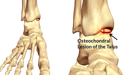
My receptionist has been complaining of left ankle pain, especially after her netball training. While treating her, I found out that her ankle injury was sustained a year ago while playing competitive netball. She's been training very hard as she's hoping to represent Singapore one day. 

She saw 3 physiotherapists concurrently last year, but none really treated her. They all just gave her exercises to do. 

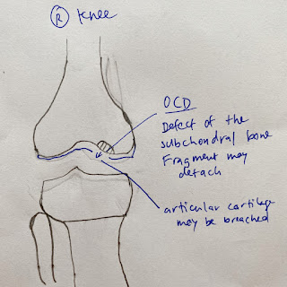
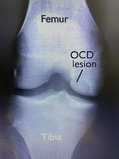
After assessing her, I told her she may have an osteochondral injury (or articular cartilage injury) in her ankle. I also shared with her a recent article (Toyooka et al, 2023) on how successful athletes are, at returning to pivoting sports after articular cartilage surgeries.

The scoping review evaluated the following articular cartilage procedures: microfractureosteochondral autograft transplanation (OAT, or mosaicplasty,  harvested from one's own joint), osteochondral allograft (OCT, using a cadaveric graft) and autologous chondrocyte implantation (ACI, or autologous chondrocyte transplantation, ACT ). All of which have been written here previously.

16 studies fulfilled the ine inclusion criteria, of which 7 studies evaluated the microfracture technique alone. 44 to 83 percent managed to return to sport (RTS) after 6.2 to 10 months. 25-75 percent managed to return to their preinjury level. Average defect size was between 1.9-4.9 cm2

87-100 percent of athletes managed to RTS after their OAT or mosaicplasty surgery after 11.8 weeks to 6.5 months.. 67-93 percent managed to get back to their preinjury levels. Mean defect size varied from 1.34 to 2.9 cm2 (this is smaller than most OAT procedures that I've read previously).

For ACI, 33-96 percent managed to RTS 10.2 months after their surgery. 26-67 percent managed to return to their preinjury levels. Mean defect size ranged from 2.1 to 6.4 cm2. These athletes had also previously undergone an average up to 2.7 other surgeries.

The rate of RTS with the microfracture technique was not higher compared to other techniques in this review. This technique is usually the first-line treatment for articular cartilage injuries since it is relatively low cost and technical ease. Patients usually RTS within 9 months. The main disadvantage is that there is no restoration of hyaline cartilage. Fibrocartilage is formed after the procedure which may not tolerate pivoting sports. It is also not suitable for those with larger defect injuries. Defect sizes larger than 2 cm2 may not have good postoperative oucomes. 

Microfracture awls to puncture holes in the bone
The OAT or mosaicplasty techniques involve harvesting a bone plug with intact cartilage from the patient's joint in a non weight bearing area and transplanting that into the defect area. The main advantage of this technique is that it has high healing potential as a patient's own bone plug is used allowing the bone to integrate immediately. There may be some risk to donor site morbidity if too big bone plugs are taken (pictured below).

Harvesting the bone plugs in the knee
87-100 percent of athletes managed to return to pivoting sports after their OAT or mosaicplasty surgery after 11.8 weeks to 6.5 months in this review. 67-93 percent managed to get back to their preinjury levels. This suggest that the OAT  procedure may offer a good acceptable result for high demand athletes. Mean defect size varied from 1.34 to 2.9 cm2 (this is smaller than most OAT procedures that I've read previously) and smallest in this review.

ACI requires 2 surgeries to restore the damages done to the hyaline cartilage lining the joint, described in more detail in a previous post. For this review, 33-96 percent managed to RTS 10.2 months after their surgery. 26-67 percent managed to return to their preinjury levels in high demand pivoting athletes. Pivoting sports may have a lower RTS ate compared with other sports.

ACI
Mean defect size ranged from 2.1 to 6.4 cm2. These athletes had also previously undergone an average up to 2.7 other surgeries. This technique is used primarily with larger defects. However, it's limitations are requring 2 surgeries, high cost, open surgery and a very prolonged rehabilitation. 

Based on this review, the OAT procedure had the highest RTS rate in pivoting sports. They also returned to sport faster, especially when the defect size is small. For large defects, OCA and ACI may be considered with ACI preferred since OCA (caderveric) has many limitations like being expensive, limited in supply and restricted in many countries. Harvested bone plugs also need to be implanted within 14-21 days.

Most studies in this review reported high RTS rates although return to preinjury level was lower. RTS is a very critical variable and benchmark (to me) for patients who are athletes. These data can be used as a basis for selecting treatment options. 

Not only there are very few studies that only study athletes, the sports they compete in also vary. There was a tendency for RTS to be higher when the level the athletes were competing in were higher (especially professional), perhaps due to their access and compliance with rehab protocols, adaptibility to competition and for financial reasons.

Note that there was not enough data on the lesion size to decide between ACI and OAT. There were no significant difference in short term results between the two, although ACI outperformed the OAT in 10 year outcomes (in 2 studies).

Well, to my receptionist and other athletes reading this post, I hope this helps with your decision making should you need to consider the different options to return to your sport. It is definitely a long and winding road, with treacherous falls along the way while attempting your comeback, but it can be done.

Trust me, I've had 3 knee surgeries, a skull fracture and broke my back twice. You just need to be be persistent and never give up. Our team in our clinics have been patients before too and know how it feels like to be a patient and will be able to understand and do their utmost to help you.


References

Bentley G, Biant LC, Vijayan S et al (2012). Minimum ten-year Results Of A Prospective Randomised Study Of Autologous Chondrocyte Implantation Versus Mosaicplasty For Symptomatic Articular Cartilage Lesions Of The Knee. JBJS Br. 94: 504-509.

Biant L, Vijayan S, Macmull S et al (2012). Autologous Chondrocyte Implantation Versus Mosaicplasty For Symptomatic Articular Cartilage Defects In The Young Adult Knee: Ten Year Results Of A Prospective Randomised Comparison Study. Orthop Proc. 94-B: 122-22

Toyooka S, Moatshe G, Persson A et al (2023). Return To Pivoting Sports After Cartilage Repair Surgery Of The Knee: A Scoping Review. Cartilage. Pub online. DOI: 10.1177/194760352211414

Monday, July 11, 2022

Viscosupplementation (Gel Shots) For Your Knees?

Picture from whichmedicaldevice.com
Many of our patients have been asked to do a hyaluronic acid injection when told that their knees were "worn out" due to osteoarthritis.

Hyaluronic acid injections (also commonly known as gel shots) are thought to help with restoring the joint fluid (synovial fluid) composition to help with lubricating and providing shock absorption to the joint. They are often suggested for patients with mild to moderate osteoarthritis to alleviate pain, reduce friction and hopefully delay surgery (total knee replacement).

The following paper is a *systematic review and meta-analysis of the largest collection of randomised trials of whether hyaluronic injections are helpful for knee osteoarthritis.

169 trials involving 21,163 patients were first identified through searches from Medline, Embase and Cochrane Register of Controlled trials. Only randomised trials with more than 100 participants per group comparing viscosupplementation with placebo or no intervention were accepted. Patients were usually given a single course of 2 to 6 injections.

Of these, 24 large, placebo controlled trials (8997 randomised patients) were included in the main analysis found that viscosupplementation (or using hyaluronic acid injections) offered a small but strong conclusive reduction in pain intensity compared with a placebo injection. 

This translated to a margin of 5 mm on a 100 mm visual analogue pain scale (or half a point on a scale of 0-10). That's a really tiny pain reduction in my opinion.

However, based on 15 large, placebo controlled trials (6463 patients), viscosupplementation was associated with a statistically significant higher risk of serious adverse events compared to a placebo.

Serious adverse events were defined as resulting in hospital admission, prolonged hospital stay, persistent or major disability, congenital abnormality of offspring, life threatening events or death.

Hence, the authors of this systematic review concluded that the findings do not support the use of viscosupplementation for the treatment of knee osteoarthritis.

Still keen on getting a hyaluronic acid injection? Mark Philippoussis (nicknamed "The Scud" after the scud missile), was a former professional tennis player known for his awesome serve. He famously used hyaluronic acid injections every 6 months for his knees to enable him to keep playing after 3 knee operations. 

But he was a former professional who was a finalist in the 1998 US Tennis Open and 2003 Wimbledon Tennis tournament.


Reference

Pereira TV, Juni P, Saadat P et al (2022). Viscusupplementation For Knee osteoarthritis: Systematic Review And Meta-analysis. BMJ. 378: e069722. DOI: 10.1136/bmj-2022-06972

*A systematic review meta analysis is a search aided by computer looking for all randomized and clinically controlled studies while a meta-analysis means using statistics to combine the data derived from a systematic review. 

Sunday, May 8, 2022

What Really Wears Out Your Joints

Picture by Dr Howard Luks
I remember treating many cases of older patients who had total hip or knee replacements due to osteoarthritis (OA) when I was a much younger physiotherapist. 

Upon asking these patients, they always wondered why they had worn out their joints despite not exercising. Some were very sedentary, while others had no time to exercise since they were more concerned with making enough to feed their families.

I've written previously that running will not wear out your knees (or your joints). So what does? There is now evidence that OA is not due to a mechanical wear and tear process. Even if you already have OA, exercise will not wear out your joints quicker. 

Of course there are mechanical causes of OA. People who are severely bow legged or have severely knocked knees can be more prone to developing OA since one side of the knee joint is over loaded. Certain fractures near a joint can lead to post traumatic OA (due to mal- alignment). 

Patients whose meniscus is torn and subsequently removed have an increased risk of developing OA. Which is why surgeons now rarely remove the whole meniscus, just the torn bit.

So, what causes arthritis in our joints? For those without any previous injuries to the joint, we have hundreds of proteins, cytokines, chemicals and other compounds that forms the articular cartilage, which lines our joints. When the joints are in good health, these chemicals support articular cartilage health and nutrition.

We do not know exactly why, whether it is due to injury, our diet, metabolism or weight that, OA develops. It may be all of the above when a 'switch' flips. Changes in the joint(s) similar to changes associated with other chronic diseases happens. That switch causes an increase the production of chemicals that harms our articular cartilage (Wang et al, 2015).

Over time, these chemicals cause injury to the articular cartilage cells. This weakens the articular cartilage and its ability to withstand load and stress.

The articular cartilage can become thinner when not functioning well. This can lead to inflammation, swelling, warmth and pain. This chronic low grade inflammation is what appears to cause OA. This is the same chronic inflammation thought to cause other chronic diseases like heart disease, fatty liver and Type II diabetes.

Researchers are still trying to understand how all these proteins and substances affect articular cartilage health and the incidences of OA. 

Exercise has actually been proven to decrease the concentration of these proteins and substances that harm our articular cartilage.

Helmark et al (2010) showed that IL-10, a chemical that protects articular cartilage in the knee was produced in response to exercise. Similarly, COMP (a protein that is a marker of cartilage degeneration) was decreased in the knee with exercise.

Another research paper by Hyldahl et al (2016) demonstrated that running was associated with a decrease in cytokines (chemicals) in the knee related to articular cartilage wear and tear. 

Studies are suggesting that metabolic health definitely plays a bigger role in causing OA. Yes, other than Type II diabetes, dementia, high blood pressure and heart disease, metabolic issues are also thought to be involved in the development of OA. All tissues in our body, including our articular cartilage are sensitive to our dietary intake. The earlier we realize this, the better off we will be.

Too many health care professionals ask their patients to stop running (or exercise) to 'save' their joints. In actual fact, running and other knee exercises have been shown to relieve mild knee arthritis and does not harm articular cartilage.

Exercise has been unequivocally proven to be the most effective treatment for early and moderate OA in our joints. You do not have to stop exercising. 

Let your symptoms be your guide. If there is no swelling and no pain, you can still run or exercise. Be careful with the distance, intensity and frequency of exercise. You may also want to cycle or swim occasionally. Or try a different shoe, or different running surface and/ or include a weight training session to get yourself stronger. 


References

Helmark IC, Mikkelsen UR, Borglum J et al (2010). Exercise Increases Interleukin-10 Levels Both Intraarticularly And Peri-synovially In patients With Knee Osteoarthritis: A Randomized Controlled Trial. Arthritis Res Ther. 12, R126. DOI: 10.1186/ar3064

Hyldahl RD, Evans A, Kwon S et al (2016). Running Decreases Knee Intra-articular Cytokine And Cartilage Oligomeric Matrix Concentrations: A Pilot Study. Eur J Appl Physiol. 116 2305-2314. DOI: 10.1007/s00421-016-3474-z

Wang X, Hunter J and Xu CD (2015). Metabolic Triggered Inflammation In Osteoarthritis. OA and Cartilage. 23(1): 22-30. DOI: 10.1016/j.joca.2014.10.002

Sunday, January 30, 2022

OCD

Kindly drawn by my older son
In case you're wondering about the title, it's not obsessive compulsive disorder, I definitely can't write a post on that. But I do know about osteochondritis dissecans (OCD). In fact when I kept having recurrent knee pain back in 2002 (which subsequently led to 3 knee operations), I initially thought I had OCD myself, before MRI scans suggested otherwise.

OCD occurs when part of a bone within a joint dies due to lack of blood flow and partially or fully separates from the joint. This separated bone piece can cause pain and subsequently restrict movement in that joint. Meaning you cannot fully straighten or bend that joint.

The cause of OCD is unknown. It is suggested that reduced blood flow to the bone may occur from repetitive trauma like excessive running and jumping. Or small, multiple episodes of minor unknown injury that damage the bone over time. Some studies suggest that there may be a genetic component involved too.

OCD occurs most frequently in children and adolescents, most commonly in the knee, but can also occur in the elbow and ankle. 

Doctors stage OCD according to how big the bone fragment is, whether the bone fragment is totally or partially separated or whether it stays in place. If that loosened piece of bone is not detached, then there may be few or no symptoms. For younger children whose bones are still growing and developing, there is a chance that it can heal.

So it was a total surprise to me when my adult patient was diagnosed with OCD 2 days ago. 

She was in the process of sitting down on her sofa and slightly twisted her knee. She could not extend or bend her knee fully nor put weight on her foot after that. She went to the hospital and was told it might be a meniscus tear and may need surgery. The surgeon suggested an MRI to confirm his hypothesis.

Hence, when the MRI results came out it was a big surprise that there was a bone fragment dislodged. That's the reason why she could not straighten or bend her knee fully.

The only incident my patient could think of was during gymnastics, when she was 12, that her knee started to hurt (possibly from the impact of repeated running and landing). Her knee would 'lock' occasionally for a few days at a time. It gradually resolved and she would occasionally have some knee pain on that same left knee while she competed in triathlons. Otherwise, there was no other clue that she would have OCD.

Now my patient has to decide how she would like to proceed to get the bone fragment out after discussion with her surgeon.


Reference

Kocher MS, Tucker R, Ganley TJ et al (2006). Management Of Osteochondritis Dissecans Of The Knee: Current Concepts Review. AJSM. 34(7): 1181-1191. DOI: 10.177/0363546506290127.

Picture from RadioGraphics 

Sunday, August 29, 2021

Locking In Your Knee May Not Mean A Meniscus Tear

Picture from ScienceDirect

A discussion with our three new physios recently revealed that they were taught that whenever a patient has a meniscal tear on MRI with mechanical symptoms it would usually mean surgery is indicated.

Mechanical symptoms are described as 'locking' or 'catching' in the knee joint that is caused by something being trapped or stuck in the knee. They were taught that it can only be removed by surgery. If the mechanical symptoms present with a tear in the meniscus, that is confirmed by MRI, it is usually attributed to the tear. Hence the rationale for surgery to remove the tear.

However, mechanical symptoms in the knee can fluctuate. Other clinicians and patients may have different variations and definitions of such 'locking' and 'catching' in the knee.

I have come across many patients with locked knees (knees that cannot fully straighten or bend), but they are seldom 'locked' all the time. We always match such symptoms in our clinic with a patient's medical or injury history and with objective orthopaedic and functional tests. 

However a study by Thorlund et al (2019) investigated whether unstable meniscal tears are more likely to cause mechanical symptoms compared to other concurrent knee pathologies like articular cartilage damage, ACL tears etc.

A wide range of meniscal tear characteristics like tear pattern, location, size of tear were included in the study. However, no important relationships were found between any of the included factors and patients reported catching or locking in the knee or inability to straighten their knees.

These results question the logic that mechanical symptoms are caused by specific joint pathologies. The authors also compared the frequency of mechanical symptoms between patients with and without a meniscal tear after knee arthroscopy. They found that half of all patients reported catching or locking. They were also unable to straighten their knee fully. However, these mechanical symptoms were equally common among patients with or without a meniscal tear.

This is consistent with my previous post where bucket handle and complex meniscal tears (both of which are commonly operated on) were found in the patient's MRI, but these patients were asymptomatic.

So if you are currently having a 'locked' knee or cannot straighten or bend your knee fully, it does not necessarily mean you need surgery. Two large scale randomized trials referenced below confirm this too. Please come and see us in our clinic for another opinion.


References

Khan M, Evaniew N, Bedi A et al (2014). Arthroscopic Surgery For Degenerative Tears Of The Meniscus: A Systematic Review And Meta-analysis. CMAJ. 186: 1057-1064

Thorlund JB, Juhl CB, Roos EM et al (2015). Arthroscopic Surgery For Degenerative Knee: A Systematic Review And Meta-analysis Of Benefits And Harms. BMJ. 350: h2747

Thorlund JB, Pihl K, Nissen N et al (2019). Conundrum Of Mechanical Knee Symptoms: Signifying Feature Of A Meniscal Tear? BJSM. 53(5): 299-303. DOI: 10.1136/bjsports-2018-09943

Sunday, May 9, 2021

Am I Crazy To Feel Pain In My Knee When My Scans Are Normal?


My patient came in yesterday saying that his knee started clicking even though he didn't feel any pain with the clicking. There were occasional twinges of pain only occasionally. However, he was worried that his knee will get worse. After assessing his knee (which turned out fine) and reassuring him, I told him my own experiences.

After I had my third knee operation (within the space of a year), my right knee started feeling better. Of course I started training again as soon as the surgeon permitted. For a start, I ran almost exclusively on grass (since it was the softest surface I could find), often going multiple rounds, to ensure that I can get my mileage since I was hoping to compete again after my injuries.  

However, I was much more sensitive about my right knee, to the point of being paranoid about every sensation I felt in the knee. Each time I was on the bus or MRT and if someone came close to my right knee, I'd move away and glare at the person for coming too close to me. Does this sound like you?

Well, it turns out this action of pain sensitization is common across other painful knee disorders as well. In patients with knee osteoarthritis, pressure from placing your hands on the knees alone can trigger pain. The good news is that this sensitization for painful knees can be treated.

The following systematic review investigated 52 studies that studied pain sensitization across four different painful knee disorders. The authors found evidence of pain sensitization in people with knee osteoarthritispatellofemoral pain and post meniscectomy patients. They however found conflicting evidence in patients with patella tendinopathy.

The researchers found that the extent of structural joint damage in the observed knee disorders does not correlate to the severity of symptoms. Hence, pain is not necessarily a 'signal' from a joint or area that is damaged.  Meaning x-rays or MRI results does not correlate with pain. Some patients have no 'damage' on x-ray/ MRI but have a lot of pain, while others with lots of damage on film may have no pain.

Many factors play a role to determine if a person will perceive a stimulus as painful or not since pain can be a complex experience that is associated with memories, belief and social context. Anxiety, depression, fear of movement, viewing their condition considerably worse than it actually is may also play a part.

It is suggested that repetitive stimulation may lead to subsequent sensitization of the nervous system. This include loading of the knee joint, ongoing inflammation at the knee joint or related tissues and altered biochemical markers. These factors contribute and maintain the pain sensitization in the knees.

In patients with knee osteoarthritis and patellofemoral joint pain, pain sensitization can be treated through exercise therapy, mobilization, pharmacological (yes, painkillers) and surgical intervention. Correct exercise is recommended for treatment of painful knees. There is the incorrect belief that exercise may harm the joint cartilage in patients with osteoarthritis.

If doctors, surgeons and physiotherapists focus less on x-rays/ MRI's and more on factors (including psychosocial factors mentioned above) relating to each patient's pain and disability, there will be more opportunities for collaboration and improved treatment outcomes.


Reference

De Oliveira Silva D, Rathleff MS, Petersen K et al (2019). Manifestations Of Pain Sensitization Across Different Painful Knee Disorders: A Systematic Review Including Meta-analysis And Metaregression. Pain Med. 20(2): 335-358. DOI: 10.1093/pm/pny177.

Saturday, November 21, 2020

Abnormal Knee MRI, But No Pain

Knee MRI by Becky Stern from Flickr
Here's a piece of surprising news for everyone. In this recently published paper I read, nearly all patients who had abnormalities on their knee 
MRI were asymptomatic, meaning no pain despite having an "abnormal" MRI.

The main inclusion criteria for this study were sedentary adults. They did not do at least 30 minutes of moderate intensity physical activity 5 days a week or 20 minutes of more intense activities 3 days a week. They did not have any knee pain, no current or previous history of knee injury and surgery.

The authors reviewed 230 knees of 115 uninjured inactive adults (51 males and 64 females). Median age was 44 years (range was 25-73 years) and all the subjects had bilateral MRI's (3.0 Tesla, high resolution) done.

Here's what they found. Brace yourself as you read on. MRI showed abnormalities in a whopping 97% of knees. 30% of knees showed tears in the meniscus. Horizontal tears were most common, while bucket handle tears least common.

Articular cartilage (57%) and bone marrow abnormalities (48%) were common in the patellofemoral (knee) joint. Moderate (19%) intensity articular cartilage lesions and severe (31%) were observed.

Grade 4 means bone rubbing on bone

Articular cartilage injuries is my area of interest since I did my postgraduate research in that area. It is interesting to note that a quarter (or 25%) of the subjects had Grade 4 changes (see picture above) visible on MRI but did not complain of pain. Perhaps this is important to remember when imaging the knee since there seems to be more visible findings here compared to the rest of the knee (articular cartilage wise).

Moderate intensity lesions were found in 21% of knee tendons while there were high grade tendonitis found in 6% of knees reviewed. The patella and quadriceps tendons being the most affected.

3% partial ligament ruptures were found, of which 2% were of the Anterior Cruciate Ligament (ACL).

The authors concluded that nearly all knees of asymptomatic adults they studied show abnormalities in at least one knee structure on MRIMeniscal tears, articular cartilage and bone marrow lesions in the patellofemoral joint were the most common pathological findings. 

They also reported finding bucket handle and complex meniscal tears (both of which commonly operated on) in asymptomatic knees. This interesting to note as bucket handle tears (as well as complex tears) would often cause 'locking' in the knee and therefore require surgery.

There you have it, the subjects were sedentary adults who did no exercise so no one can say that it was running or exercise that "wore out" their joints. And some these very same adults had "terrible" or abnormal MRI's, but were asymptomatic or did not have any pain.

So don't fret if your MRI is abnormal. You may not need any surgical intervention, especially if you do not have any pain or if that pain is easily treated.

Maybe these abnormalities should be just described as "wrinkles" on the inside. 


Reference

Horga LM, Hirschman AC, Henckel J et al (2020). Prevalence Of Abnormal Findings In 230 Knees Of Asymptomatic Adults Using 3.0 T MRI. Skele Radiol 49: 1099-1107. DOI: 10.1007/s00256-020-03394-z

Sunday, March 24, 2019

Still Can't Run 6 Months After An Ankle Sprain


R ankle
My last patient yesterday suffered an osteochondral injury in her right ankle last July after spraining her ankle while taking part in a trail running camp. Part of the reason may be wearing a new pair of shoes she wasn't used to.

Almost half a year later, she was still hoping to do a 50 km trail race earlier this month, but she definitely wasn't even ready to be running yet.

Many of her running friends and colleagues (she works in a hospital) can't believe an osteochondral injury can be that serious. In fact, some of the physiotherapists in her hospital don't even know what an osteochondral injury to her talus means. They definitely do not know how to treat her.

Hence, you have a runner who works in a hospital, yet comes to Sports Solutions to get treatment.  Just like the physiotherapist from another hospital who sees us after her microfracture surgery.

In the picture above, you can see where the talus is in the ankle. It is the bone that is below your tibial (shin bone). In the scenario where there is an injury to the articular cartilage (which lines the end of our bones to allow for load bearing and friction free movement), there will be both swelling and pain.

The main goal of non operative treatment is to allow the injured bone and articular cartilage to heal. Since articular cartilage has poor blood supply, this is not going to happen quickly.

Sometimes, crutches or a rocker boot may be necessary to take load off weight bearing. In severe cases, surgery is needed to ensure recovery. After three articles on the operative management on articular cartilage injuries, I think I've covered the topic enough. Just in case you're keen, drilling can be done like the picture you see below to stimulate healing.
Drilling is done
I've told my patient she can still volunteer at the race whether it's manning a water station, handing out medals or directing runners at checkpoints in the race. She can also lend support and cheer for her friends at the race. Meanwhile, she has also found "a distraction" or another way to motivate herself. She's been going to the gym regularly and her upper body is much more muscular than before.

I've told her to keep the long view in mind. If she wants to keep running for the rest of her life it's not worth risking it now. There will be other races for her in future definitely.

Have an osteochondral injury? Come see us in our clinics.

Sunday, March 10, 2019

Autologous Chondrocyte Transplantation For Articular Cartilage Injuries


In the two previous posts, we discussed the microfracture technique and the mosaicplasty technique for articular injuries. The third major procedure for articular cartilage injuries is autologous chondrocyte transplanatation (ACT) or autologous chondrocyte implantation (ACI). This is also the most invasive of the three. This method is usually chosen if the defect is larger than 5 cm and especially if there's a "kissing lesion" (or touching lesions on two joint surfaces).
Kissing lesions- both surfaces affected
The patient undergoes two surgeries for this. In the first, a small patch of healthy articular cartilage the size of one of two Tic Tacs is harvested (from the knee) and sent to a laboratory. It is subsequently grown in a protected medium to get more healthy articular cartilage. During the second surgery two to three weeks later, these newly grown articular cartilage cells are placed onto the defect (which is cleaned) to restore the surface.


There are of course variations to the three surgical interventions described in these few posts like a cell based, biodegradeable membarnes or scaffolding, stem cells etc.

Return to light sporting activities is usually allowed after six months with full return to sports at around nine to twelve months after the second surgery depending on how the patient recovers.

These have strong implications for physiotherapists in the management of these disorders as physiotherapists take charge of the patient's rehabilitation program after surgery. Successful rehabilitation for a patient requires the physiotherapist to have knowledge of the biology of articular cartilage and the factors that will influence damage and repair.

This requires restoring motion and muscle function while reducing functional limitations during weight bearing activities. Patient education and setting of realistic goals based on the extent of the damage is crucial to a successful outcome.

The postoperative management of patient varies according to the surgery performed. There are different time frames for non and partial weight bearing, specific physiotherapy treatment and use of continuous passive motion (CPM) machines. Other than improving range, CPM machines provide a mechanical stimulus to joints to promote healing (Sledge, 2001).

A good surgical technique is only as good as its rehabilitation. Come and see us if you have articular cartilage injuries as we definitely know what to do.


References

Brittberg M, Lindahl A et al (1994). N Eng J Med. 331(14): 889-95. DOI: 10.1056/NEJM199410063311401.

Vasiliadis HS and Wasiak J (2016). Autologous Chondrocyte Implantation For Full Thickness Articular Cartilage Defects Of The Knee. Cochrane Database of Systematic Reviews. Issue 10. Art. No CD003323. DOI: 10.1002/14651858.CD003323.pub3.

Sledge, SL (2001). Microfracture Techniques In The Treatment Of Osteochondral Injuries. Clinics Sp Med. 20(2): 365-377.

Sunday, March 3, 2019

Mosaicplasty For Articular Cartilage Injuries


Last week we discussed the microfracture technique for articular cartilage injuries. Many of my patients who read the article commented that they did not realize it was such a serious condition. Yes, indeed, having an articular cartilage injury is worse than tearing your anterior cruciate ligament (ACL). The lengthy rehabilitation makes it much worse.

Generally, the microfracture technique works well only for the smaller lesions (less than 3 cm). If the size of the defect is larger, the surgeon would usually perform mosaicplasty or autologous chondrocyte transplantation (ACT). The latter procedure works better if there is a "kissing lesion" (defect on both joint surfaces).

I wrote that the microfracture technique was made popular by Richard Steadman. For mosaicplasty, it is Professor Laszlo Hangody from Hungary who has probably performed the most procedures and published the most articles on the topic.
Harvesting the bone plugs

Mosaicplasty is a technique in which small bone plugs with healthy hyaline cartilage are taken and then transplanted to cover the defect in the damaged area. The bone plugs are usually taken from an area that is non weight bearing to cover the defect in the affected area. The end result ends up looking like mosaic tiles, hence the name mosaicplasty.
Here's a closer look

The hope is that the body will not miss the taken parts and it can be used where it is needed. Over time, the holes in that part of  bone that is taken will fill with bone and scar tissue. The bone plugs can be from the patient or from fresh cadavers.

The above diagram shows the procedure done on a patient's knee, but it can be done on the ankle, hip and other weight bearing articular surfaces too.


A similar technique to mosaicplasty is Osteochondral Autograft Transfer System or (OATS). The bone plugs used in OATS are usually larger and usually only one or two plugs are needed to fill the area of damage.
See the gaps in between the bone plugs?
In the above pictures, you can see that there are still gaps between the cylindrical bone plugs. This is the main problem with this particular technique. The defect is not filled completely and the gaps normally fill up with fibrocartilage. There is then a worry about how this holds up over time, especially if the patient is keen on returning to sports.


Recently, there has been an improvement/ modification to this technique. A group of researchers, inspired by the honey comb structure of a beehive of honey bees decided to use hexagonal shaped bone plugs instead of the cylindrical ones used previously.
No more gaps?
This is to eliminate the gaps while performing the procedure. The authors named it hexagonal osteochondral graft system (HOGS). Early outcomes of HOGS seemed comparable to mosaicplasty and promising at this stage although further follow up needs to be done.

Advantages of mosaicplasty  are that only one operation is needed (compared to two in ACT) and hence lower cost and less down time. There is less risk of disease transmission and there is a high percentage of hyaline cartilage for the damaged surface.

These procedures require the physiotherapist treating the patient to have knowledge of the biology of articular cartilage and the factors that may influence degradation and repair. The physiotherapist needs to know the nature, location, size of lesion and the surgical procedure performed.

Rehabilitation should address the patient's impairments and functional limitations without jeopardizing healing of the lesion.

I remember back in 1999, as a young physiotherapist a patient told me he had mosaicplasty done and I had asked him what it was. That piqued my interest in articular cartilage injuries and especially so when I later had to have a microfracture procedure done on my right knee in 2003. That also was why I did postgraduate work in that area as I desperately wanted to compete again.

My next post will be on the autologous chondrocyte transplantation/ implantation (ACT) procedure.

References

Erol MF and Karakoyun O. (2016). A New Point Of View For Mosaicplasty In The Treatment Of Focal Cartilage Defects Of Knee Joint: Honeycomb Pattern. SpringerPlus. 5(1): 1170. DOI: 10.1186/s40064-016-2796-y.

Gracitelli GC, Moraes VY et al (2016). Surgical Interventions (Microfracture, Drilling, Mosaicplasty And Allograft Transplantation) For Treating Isolated Cartilage Defects Of The Knee In Adults. Cochrane Database of Systematic Reviews. Issue 9. Art. No CD10675. DOI: 10.1002/4651858.CD010675.pub2.

Hangody L and Balo E (2011). Autologous Osteochondral Mosaicplasty. In Sanchis-Alfonso V. (eds). Anterior Knee Pain And Patellar Instability, London.

Sunday, February 24, 2019

My Patient Had A Microfracture Done In The Knee

Articular cartilage is white, with the bone exposed
A fellow physiotherapist who works in a hospital came to see me after a microfracture procedure done on her knee. There was a grade 4 articular cartilage defect on her knee and this led to swelling and pain while climbing stairs and after weight training.

Outerbridge articular cartilage classification
All of us have a layer of articular cartilage covering the ends of  our bones, especially the joint surfaces. It is normally tough and resilient. This helps to protect the joint during load bearing and reduce friction during movement. Injury or damage to the articular cartilage can result from trauma (during sports) or from daily wear and tear. As articular cartilage has poor/ no blood supply, it does not heal well after injury.

There is no standard and uniform approach to managing articular cartilage injuries in the knee. Left untreated it can progress to significant joint destruction. The patient may then need a total knee replacement in the worse case scenario.

Treatment options include microfracture, arthoscopic drilling, mosaicplasty and chondrocyte transplantation to restore the joint surface.

A microfracture technique is where the surgeon performs key hole surgery to cause bleeding on the bone surface to promote healing (picture below). It is performed by the surgeon puncturing holes in the subchondral bone layer to allow bleeding to occur.  After the blood clots and heals, a layer of fibrocartilage is formed. This technique was first made popular more than 20 years ago by Dr Richard Steadman from Vail, Colorado who has since retired.

My patient had the microfracture procedure done (in the picture above) and as you can imagine, the rehabilitation to return to sport can be lengthy. There is usually a period of non weight bearing for the first six to eight weeks to allow healing while using continuous passive motion (CPM)  machine at night. Use of the CPM machine is to stimulate movement to enable nutrition in the articular cartilage since the patient is non weight bearing. Yes, correct movement and some loading forces are necessary for our articular cartilage to recuperate.

Most surgeons here do not usually suggest use of the CPM machine after performing the microfracture technique which I feel is critical in order for optimal healing to occur in the articular cartilage.

Lots of patience and consistency are required by the patient and physiotherapist to slowly regain functional range of movement and strength before any return to sport work can be done.

Fortunately, articular cartilage injuries are my area of interest having had them myself (and requiring 3 knee surgeries) and my postgraduate research was in this area.

I'll write more about mosaicplasty and and the chondrocyte transplantation procedure in the next post. Stay tuned.


References

Hurst JM, Steadman JR et al (2010). Rehabilitation Following Microfracture For Chondral Injury In The Knee. Clin Sports Med. 29(2): 257-265. DOI: 10.1016/j.csm.2009012.009.

Steadmann JR, Rodkey WG et al (1997). Microfracture Technique For Full-thickness Chondral Defects. Oper Tech Orthop. 7:300-304.

Here's what the surgeons use to cause bleeding in the bone

Friday, August 10, 2018

Fat Pad Most Painful In The Knee?

I had a patient who came to our clinic recently complaining that his MRI showed that his patella (knee cap) cartilage had "worn out" completely but he didn't have any pain prior to that. He had actually gone to do his MRI under his doctor's insistence for investigating something else.

His  MRI results was like in his words "opening a can of worms" telling him what's wrong with his knees and perhaps that's why he started having pain after that.

After his ranting, I had to explain very thoroughly about the structures in our knees that cause the most pain. The information I gave him was derived from an article published quite a while ago in the American Journal of Sports Medicine but still very relevant today.

The doctors in that study came up with a simple method to document the various sensations felt inside a single subject's knees one week apart. Right knee first, followed by the left a week later. (Note that the subject had no prior knee pain).

They would arthroscopically poke/ palpate (using a specially built spring loaded device) different structures inside the knee while video recording the procedure and record what the subject's response was. Force used was between 0 to 500 grams. All this done without intra articular anesthesia. Ouch! That must really hurt.

The doctors only injected local anesthesia at the portal site (incision). The first author inspected both knees arthroscopically. He asked the patient when he poked at different structures and graded the sensation as follows (0) no sensation; (1) was non painful awareness; (2) slight discomfort; (3) moderate discomfort and (4) severe pain. This was done with with a modifier of either accurate spatial localization (A) or poor spatial localization (B).

Ready for the results? They were exactly the same for both knees. Even though it was done one week apart.

Palpation of the patellar articular cartilage in the three under surfaces (central ridge, medial and lateral facets) resulted in no sensation, or a 0 score, even with a strongest force of 500 grams. Palpation of the odd facets elicited a score of 1B. Asymptomatic grade II or III chondromalacia (wearing out) of the central ridge was identified on both patellas of the subject!

Palpation of the articular cartilage surfaces of the femoral condyles, trochlea, and tibial plateaus at 500 g of force universally produced a sensation of 1B to 2B.

The sensation from the meniscus ranged from 1B on the inner rim of the meniscus to to 3B near the capsular margin.

Sensation from the  cruciate liagaments (Anterior, posterior cruciate ligaments) range from 1-2B in the mid-portion of the ligaments and 3-4B at the insertion sites.

Palpation of the suprapatellar pouch, capsule, and the medial and lateral retinacula produced a score of 3A to 4A (moderate to severe localized pain) at relatively low levels of force (about 100 g).


The most painful structures were the anterior synovium of the knee, the fat pad and the joint capsule - 4A.

The human knee can be very complex, especially our patellofemoral joint (patella and the femur). The three asymnetrical surfaces on the underside of the patella (or knee cap) has to work together with the femur as it accepts, transfers and dissipates loads between the bones.

We know from previous research that various structures in the knee send neurosensory signals (or messages) to the brain. It is theses signals that result in us feeling pain.

Even though my patient's patella cartilage had worn out (just like the subject) there shouldn't be any pain there as articular cartilage doesn't have any nerve supply. No nerve endings means it is unable to detect pain.

Even the ACL and meniscus wasn't really that sensitive to the poking. This observation may provide an explanation for the often poor localization of structural damage that many patients experience with a cruciate ligament or meniscal injury.

Now you know, worn out articular cartilage doesn't cause you pain. The pain you have is likely to come from other structures. And you definitely don't need to ingest any glucosamine too.


Reference

SF Dye, GL Vaupel and CC Dye (1998). Conscious Neurosensory Mapping Of The Internal Structures Of The Human Knee Without Intraarticular Anesthesia. AM J Sp Med. 26(6): 773-777. DOI: 10.1177/03635465980260060601.
black and white version