Showing posts sorted by relevance for query osteochondral. Sort by date Show all posts

Sunday, March 24, 2019

Still Can't Run 6 Months After An Ankle Sprain


R ankle
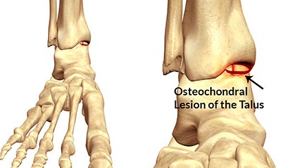
My last patient yesterday suffered an osteochondral injury in her right ankle last July after spraining her ankle while taking part in a trail running camp. Part of the reason may be wearing a new pair of shoes she wasn't used to.

Almost half a year later, she was still hoping to do a 50 km trail race earlier this month, but she definitely wasn't even ready to be running yet.

Many of her running friends and colleagues (she works in a hospital) can't believe an osteochondral injury can be that serious. In fact, some of the physiotherapists in her hospital don't even know what an osteochondral injury to her talus means. They definitely do not know how to treat her.

Hence, you have a runner who works in a hospital, yet comes to Sports Solutions to get treatment.  Just like the physiotherapist from another hospital who sees us after her microfracture surgery.

In the picture above, you can see where the talus is in the ankle. It is the bone that is below your tibial (shin bone). In the scenario where there is an injury to the articular cartilage (which lines the end of our bones to allow for load bearing and friction free movement), there will be both swelling and pain.

The main goal of non operative treatment is to allow the injured bone and articular cartilage to heal. Since articular cartilage has poor blood supply, this is not going to happen quickly.

Sometimes, crutches or a rocker boot may be necessary to take load off weight bearing. In severe cases, surgery is needed to ensure recovery. After three articles on the operative management on articular cartilage injuries, I think I've covered the topic enough. Just in case you're keen, drilling can be done like the picture you see below to stimulate healing.
Drilling is done
I've told my patient she can still volunteer at the race whether it's manning a water station, handing out medals or directing runners at checkpoints in the race. She can also lend support and cheer for her friends at the race. Meanwhile, she has also found "a distraction" or another way to motivate herself. She's been going to the gym regularly and her upper body is much more muscular than before.

I've told her to keep the long view in mind. If she wants to keep running for the rest of her life it's not worth risking it now. There will be other races for her in future definitely.

Have an osteochondral injury? Come see us in our clinics.

Sunday, February 26, 2023

Is It Easy To Return To Pivoting Sports After Articular Cartilage Surgery?

Picture from Upswinghealth
My receptionist has been complaining of left ankle pain, especially after her netball training. While treating her, I found out that her ankle injury was sustained a year ago while playing competitive netball. She's been training very hard as she's hoping to represent Singapore one day. 

She saw 3 physiotherapists concurrently last year, but none really treated her. They all just gave her exercises to do. 

After assessing her, I told her she may have an osteochondral injury (or articular cartilage injury) in her ankle. I also shared with her a recent article (Toyooka et al, 2023) on how successful athletes are, at returning to pivoting sports after articular cartilage surgeries.

The scoping review evaluated the following articular cartilage procedures: microfractureosteochondral autograft transplanation (OAT, or mosaicplasty,  harvested from one's own joint), osteochondral allograft (OCT, using a cadaveric graft) and autologous chondrocyte implantation (ACI, or autologous chondrocyte transplantation, ACT ). All of which have been written here previously.

16 studies fulfilled the ine inclusion criteria, of which 7 studies evaluated the microfracture technique alone. 44 to 83 percent managed to return to sport (RTS) after 6.2 to 10 months. 25-75 percent managed to return to their preinjury level. Average defect size was between 1.9-4.9 cm2

87-100 percent of athletes managed to RTS after their OAT or mosaicplasty surgery after 11.8 weeks to 6.5 months.. 67-93 percent managed to get back to their preinjury levels. Mean defect size varied from 1.34 to 2.9 cm2 (this is smaller than most OAT procedures that I've read previously).

For ACI, 33-96 percent managed to RTS 10.2 months after their surgery. 26-67 percent managed to return to their preinjury levels. Mean defect size ranged from 2.1 to 6.4 cm2. These athletes had also previously undergone an average up to 2.7 other surgeries.

The rate of RTS with the microfracture technique was not higher compared to other techniques in this review. This technique is usually the first-line treatment for articular cartilage injuries since it is relatively low cost and technical ease. Patients usually RTS within 9 months. The main disadvantage is that there is no restoration of hyaline cartilage. Fibrocartilage is formed after the procedure which may not tolerate pivoting sports. It is also not suitable for those with larger defect injuries. Defect sizes larger than 2 cm2 may not have good postoperative oucomes. 

Microfracture awls to puncture holes in the bone
The OAT or mosaicplasty techniques involve harvesting a bone plug with intact cartilage from the patient's joint in a non weight bearing area and transplanting that into the defect area. The main advantage of this technique is that it has high healing potential as a patient's own bone plug is used allowing the bone to integrate immediately. There may be some risk to donor site morbidity if too big bone plugs are taken (pictured below).

Harvesting the bone plugs in the knee
87-100 percent of athletes managed to return to pivoting sports after their OAT or mosaicplasty surgery after 11.8 weeks to 6.5 months in this review. 67-93 percent managed to get back to their preinjury levels. This suggest that the OAT  procedure may offer a good acceptable result for high demand athletes. Mean defect size varied from 1.34 to 2.9 cm2 (this is smaller than most OAT procedures that I've read previously) and smallest in this review.

ACI requires 2 surgeries to restore the damages done to the hyaline cartilage lining the joint, described in more detail in a previous post. For this review, 33-96 percent managed to RTS 10.2 months after their surgery. 26-67 percent managed to return to their preinjury levels in high demand pivoting athletes. Pivoting sports may have a lower RTS ate compared with other sports.

ACI
Mean defect size ranged from 2.1 to 6.4 cm2. These athletes had also previously undergone an average up to 2.7 other surgeries. This technique is used primarily with larger defects. However, it's limitations are requring 2 surgeries, high cost, open surgery and a very prolonged rehabilitation. 

Based on this review, the OAT procedure had the highest RTS rate in pivoting sports. They also returned to sport faster, especially when the defect size is small. For large defects, OCA and ACI may be considered with ACI preferred since OCA (caderveric) has many limitations like being expensive, limited in supply and restricted in many countries. Harvested bone plugs also need to be implanted within 14-21 days.

Most studies in this review reported high RTS rates although return to preinjury level was lower. RTS is a very critical variable and benchmark (to me) for patients who are athletes. These data can be used as a basis for selecting treatment options. 

Not only there are very few studies that only study athletes, the sports they compete in also vary. There was a tendency for RTS to be higher when the level the athletes were competing in were higher (especially professional), perhaps due to their access and compliance with rehab protocols, adaptibility to competition and for financial reasons.

Note that there was not enough data on the lesion size to decide between ACI and OAT. There were no significant difference in short term results between the two, although ACI outperformed the OAT in 10 year outcomes (in 2 studies).

Well, to my receptionist and other athletes reading this post, I hope this helps with your decision making should you need to consider the different options to return to your sport. It is definitely a long and winding road, with treacherous falls along the way while attempting your comeback, but it can be done.

Trust me, I've had 3 knee surgeries, a skull fracture and broke my back twice. You just need to be be persistent and never give up. Our team in our clinics have been patients before too and know how it feels like to be a patient and will be able to understand and do their utmost to help you.


References

Bentley G, Biant LC, Vijayan S et al (2012). Minimum ten-year Results Of A Prospective Randomised Study Of Autologous Chondrocyte Implantation Versus Mosaicplasty For Symptomatic Articular Cartilage Lesions Of The Knee. JBJS Br. 94: 504-509.

Biant L, Vijayan S, Macmull S et al (2012). Autologous Chondrocyte Implantation Versus Mosaicplasty For Symptomatic Articular Cartilage Defects In The Young Adult Knee: Ten Year Results Of A Prospective Randomised Comparison Study. Orthop Proc. 94-B: 122-22

Toyooka S, Moatshe G, Persson A et al (2023). Return To Pivoting Sports After Cartilage Repair Surgery Of The Knee: A Scoping Review. Cartilage. Pub online. DOI: 10.1177/194760352211414

Sunday, March 3, 2019

Mosaicplasty For Articular Cartilage Injuries


Last week we discussed the microfracture technique for articular cartilage injuries. Many of my patients who read the article commented that they did not realize it was such a serious condition. Yes, indeed, having an articular cartilage injury is worse than tearing your anterior cruciate ligament (ACL). The lengthy rehabilitation makes it much worse.

Generally, the microfracture technique works well only for the smaller lesions (less than 3 cm). If the size of the defect is larger, the surgeon would usually perform mosaicplasty or autologous chondrocyte transplantation (ACT). The latter procedure works better if there is a "kissing lesion" (defect on both joint surfaces).

I wrote that the microfracture technique was made popular by Richard Steadman. For mosaicplasty, it is Professor Laszlo Hangody from Hungary who has probably performed the most procedures and published the most articles on the topic.
Harvesting the bone plugs

Mosaicplasty is a technique in which small bone plugs with healthy hyaline cartilage are taken and then transplanted to cover the defect in the damaged area. The bone plugs are usually taken from an area that is non weight bearing to cover the defect in the affected area. The end result ends up looking like mosaic tiles, hence the name mosaicplasty.
Here's a closer look

The hope is that the body will not miss the taken parts and it can be used where it is needed. Over time, the holes in that part of  bone that is taken will fill with bone and scar tissue. The bone plugs can be from the patient or from fresh cadavers.

The above diagram shows the procedure done on a patient's knee, but it can be done on the ankle, hip and other weight bearing articular surfaces too.


A similar technique to mosaicplasty is Osteochondral Autograft Transfer System or (OATS). The bone plugs used in OATS are usually larger and usually only one or two plugs are needed to fill the area of damage.
See the gaps in between the bone plugs?
In the above pictures, you can see that there are still gaps between the cylindrical bone plugs. This is the main problem with this particular technique. The defect is not filled completely and the gaps normally fill up with fibrocartilage. There is then a worry about how this holds up over time, especially if the patient is keen on returning to sports.


Recently, there has been an improvement/ modification to this technique. A group of researchers, inspired by the honey comb structure of a beehive of honey bees decided to use hexagonal shaped bone plugs instead of the cylindrical ones used previously.
No more gaps?
This is to eliminate the gaps while performing the procedure. The authors named it hexagonal osteochondral graft system (HOGS). Early outcomes of HOGS seemed comparable to mosaicplasty and promising at this stage although further follow up needs to be done.

Advantages of mosaicplasty  are that only one operation is needed (compared to two in ACT) and hence lower cost and less down time. There is less risk of disease transmission and there is a high percentage of hyaline cartilage for the damaged surface.

These procedures require the physiotherapist treating the patient to have knowledge of the biology of articular cartilage and the factors that may influence degradation and repair. The physiotherapist needs to know the nature, location, size of lesion and the surgical procedure performed.

Rehabilitation should address the patient's impairments and functional limitations without jeopardizing healing of the lesion.

I remember back in 1999, as a young physiotherapist a patient told me he had mosaicplasty done and I had asked him what it was. That piqued my interest in articular cartilage injuries and especially so when I later had to have a microfracture procedure done on my right knee in 2003. That also was why I did postgraduate work in that area as I desperately wanted to compete again.

My next post will be on the autologous chondrocyte transplantation/ implantation (ACT) procedure.

References

Erol MF and Karakoyun O. (2016). A New Point Of View For Mosaicplasty In The Treatment Of Focal Cartilage Defects Of Knee Joint: Honeycomb Pattern. SpringerPlus. 5(1): 1170. DOI: 10.1186/s40064-016-2796-y.

Gracitelli GC, Moraes VY et al (2016). Surgical Interventions (Microfracture, Drilling, Mosaicplasty And Allograft Transplantation) For Treating Isolated Cartilage Defects Of The Knee In Adults. Cochrane Database of Systematic Reviews. Issue 9. Art. No CD10675. DOI: 10.1002/4651858.CD010675.pub2.

Hangody L and Balo E (2011). Autologous Osteochondral Mosaicplasty. In Sanchis-Alfonso V. (eds). Anterior Knee Pain And Patellar Instability, London.

Sunday, March 10, 2019

Autologous Chondrocyte Transplantation For Articular Cartilage Injuries


In the two previous posts, we discussed the microfracture technique and the mosaicplasty technique for articular injuries. The third major procedure for articular cartilage injuries is autologous chondrocyte transplanatation (ACT) or autologous chondrocyte implantation (ACI). This is also the most invasive of the three. This method is usually chosen if the defect is larger than 5 cm and especially if there's a "kissing lesion" (or touching lesions on two joint surfaces).
Kissing lesions- both surfaces affected
The patient undergoes two surgeries for this. In the first, a small patch of healthy articular cartilage the size of one of two Tic Tacs is harvested (from the knee) and sent to a laboratory. It is subsequently grown in a protected medium to get more healthy articular cartilage. During the second surgery two to three weeks later, these newly grown articular cartilage cells are placed onto the defect (which is cleaned) to restore the surface.


There are of course variations to the three surgical interventions described in these few posts like a cell based, biodegradeable membarnes or scaffolding, stem cells etc.

Return to light sporting activities is usually allowed after six months with full return to sports at around nine to twelve months after the second surgery depending on how the patient recovers.

These have strong implications for physiotherapists in the management of these disorders as physiotherapists take charge of the patient's rehabilitation program after surgery. Successful rehabilitation for a patient requires the physiotherapist to have knowledge of the biology of articular cartilage and the factors that will influence damage and repair.

This requires restoring motion and muscle function while reducing functional limitations during weight bearing activities. Patient education and setting of realistic goals based on the extent of the damage is crucial to a successful outcome.

The postoperative management of patient varies according to the surgery performed. There are different time frames for non and partial weight bearing, specific physiotherapy treatment and use of continuous passive motion (CPM) machines. Other than improving range, CPM machines provide a mechanical stimulus to joints to promote healing (Sledge, 2001).

A good surgical technique is only as good as its rehabilitation. Come and see us if you have articular cartilage injuries as we definitely know what to do.


References

Brittberg M, Lindahl A et al (1994). N Eng J Med. 331(14): 889-95. DOI: 10.1056/NEJM199410063311401.

Vasiliadis HS and Wasiak J (2016). Autologous Chondrocyte Implantation For Full Thickness Articular Cartilage Defects Of The Knee. Cochrane Database of Systematic Reviews. Issue 10. Art. No CD003323. DOI: 10.1002/14651858.CD003323.pub3.

Sledge, SL (2001). Microfracture Techniques In The Treatment Of Osteochondral Injuries. Clinics Sp Med. 20(2): 365-377.