Showing posts with label Low back pain. Show all posts

Sunday, May 11, 2025

Is The Adductor Magnus Muscle Really An Adductor?

Picture from Getbodysmart
Our adductor magnus muscle is a very large muscle in the medial (inner) part of the thigh. It has long been thought to be a hip adductor. If you are standing with your feet wide apart and you bring your left leg in towards the midline, you will be adducting your left leg. 

Think about that movement, we do not always actively adduct a lot do we? Even while walking or running, we do not need to adduct much. Why is the adductor magnus muscle so big if we do not adduct a lot? Unless you ride horses, donkeys or ponys, what do we need such large hip adductors for?

Adductor magnus also helps with flexion and medial rotation of the hip. And if you were to read about group of Japanese researchers work, it is actually a very strong hip extensor. 

A group of Japanese researchers (Takahashi et al, 2025) tested the hypothesis that the adductor magnus is actually a hip extensor more than a hip adductor.

Picture from Takahashi et al, 2025
The researchers utilized advanced diffusion tensor imaging and reconstructed the entire muscle in 15 young adults pictured above. Adductor magnus is divided into 3 portions based on fascicle insertion. The posterior (back) and anterior-distal portions comprised over 80 percent of the whole muscle volume and cross-sectional area. These 2 portions demonstrated that hip extension was more commonly being activated rather than hip adduction.

Because of this, the maximal force generating capacity of the whole muscle was over 2 fold greater for hip extension than adduction. These results support the authors' hypothesis that adductor magnus is actually a major hip extensor rather than hip adductor, challenging the traditional view of this muscle as a hip adductor.

Those of you who have read this far (thank you) must be wondering what is the big deal? Or how does this help? Consider the following pictures that show a more 3D view of the adductor magnus.

Picture by John Hull Grundy
Look at the right leg in the picture on the left. The top part is actually adductor longus. See the twist below rarely seen in 2 dimensional anatomy books? This twist allows adductor magnus to be a major stabilizer of the pelvis (hip). If you bend down to pick something off the ground, the large muscles on the front, side and back of your pelvis are doing most of the work while adductor magnus prevents them working together to throw you off balance. It is uniquely positioned to resist too much hip flexion, extension, lateral rotation of the hip, swaying side to side and hip abduction. 

Many therapists may not know that adductor magnus' squarish shape and twisted nature makes it a very important pelvic stabilizer. One that we cannot ignore when patients come in with hip or back pain.

Yes back pain included. The longest part of adductor magnus is at the back (pictured above from Anatomy Trains). It almost looks like a separate muscle with its fibers going straight down from the ischial tuberosity (just like the hamstrings) and finishing at the medial epicondyle of the femur (inner part of the knee).
 
This part of adductor magnus keeps the pelvis and your upper body from falling forward. This is done much more efficiently by this part of adductor magnus compared to the hamstrings. So when patients or other therapists tell you that you have "tight" or "short" hamstrings, it is because your hamstrings are working too hard to stabilize your pelvis and upper body from falling forward.

The next time you have low back pain or hamstring pain, consider getting your adductor magnus checked!

Reference

Takahashi K, Tozawa H, Kawama R et al 92025). Redefining Muscular Action: Human "Adductor" Magnus Is Designed To Act Primarily For Hip "Extension" Rather Than Adduction In Young Living Individuals. J App Physiol. DOI: 10.1152/japplphysiol.00600.2024

Sunday, June 9, 2024

Understanding The Recovery Of The Intervertebral Disc

Picture from Wikipedia
I came across a recent paper (Feki et al, 2024) about the positions which help our intervertebral discs recover and heal when we sleep. I have written at least 5 articles on intervertebral discs. This is definitely still a topic that I get questions from most often among my friends and patients alike. Almost every single one of my friends and patients are fearful of having a 'slipped intervertebral disc' whenever they have low back pain (LBP). The intervertebral discs (IVD) have a really bad reputation for causing significant pain and disability in many people.

Here's a quick review if you did not read those 5 articles.


The IVD consists of a very tough outer layer called the annulus fibrosis (AF). It is made of of several layers of fibrocartilage consisting of Type I and II collagen fibers. The AF protects the soft, gel-like substance in the middle known as the nucleus pulposus (NP). The NP helps distribute pressure evenly across the IVD and prevent excessive forces on the spine.


See how thick the AF is from the picture above? Here's something else you need to know. There is a cartilaginous endplate between the AF and the vertebra (the spine). The endplates hold the IVD in place. It allows load to be spread evenly and to provide attachment to the IVD. This creates a super strong connection to the AF making it impossible for the IVD to 'slip' out of position.

Yes, our discs are actually very strong and hardy. How strong are our IVD's? In a published study on thoracic discs in the young (28 years old plus minus 8 years) , it took about 740 pounds of force to compress the disc height 1 mm. For the older subjects (70 years young plus minus 7 years), it took almost 460 pounds of force. Note that these are on cadavers with the muscles and bones cut away (Stemper et al, 2010).

The IVD's withstand a whole variety of complex forces in our daily activities and also when we exercise. This can lead to significant structural changes in terms of volume, area and height of the intervertebral disc. It can lead to an increase in disc stiffness and a decrease in interdiscal pressure.

In order for your discs to remain healthy and strong, it is dependent on a recovery phase which serves to prevent premature disc degeneration. This happens during a period of nocturnal rest i.e. when you sleep at night.

This phenomenon of disc recovery has been documented extensively through many studies using MRI and intradiscal pressure measurement. Fluid dynamics (water content) within the disc are considered a primary factor in recovery, while it's intricate multiscale structure and viscoelastic (behaving with both liquid-like and solid-like) properties also play key roles.

Feki et al (2024) in their review collated, analyzed and evaluated the existing in vivo (human) and in vitro (in controlled environments) on this topic to provide a comprehensive understanding of this recovery process to enable future advancements in medical treatment and biomedical enginerring solutions to enhance the natural recovery processes of intervertebral discs.

As it is a very long (and complex) review article (Feki et al, 2024), I am simply highlighting the optimal recovery positions (pictured below) for intervertebral disc rehydration. 
Picture from Feki et al, 2024
(a) prone (b) modified press up/ push up position (c) supine with under knee and back support
(d) and (e) side lying with lumbar flexion with pillows between legs
(f) and (g) side lying with and without manual distraction
(h) 50 degrees gravity assisted position (i) 110 degrees supported sitting
(j) inclined sitting with lumbar support

So, you see from the pictures that some of the supposedly 'poor' posture/ positions you were told, are actually good for your intervertebral discs.

References

Feki F, Zairi F, Tamoud A et al (2024). Understanding The Recovery Of The Intervertebral Disc: A Comprehensive Review Of In Vivo And In Vitro Studies. J Bionic Eng. DOI: 10.1007/s42235-024-00542-2

Fournier DE, Kiser PK, Shoemaker JK et al (2020). Vascularization Of The Human Intervertebral Disc: A Scoping Review. JOR Spine. 15: 3(4): e1123. DOI: 10.1002/jsp2.1123.

Stemper BD, Board D et al (2010). Biomechanical Properties Of Human Thoracic Spine Disc Segments. J Craniovert Junct Sp. 1(1): 18-22. DOI: 10.4103/09774-8237.65477

Sunday, September 4, 2022

The Larger The Disc Injury The More Likely To Heal Without Surgery


Picture from article referenced below

I saw a few patients with slipped discs this past week. Most patients are still very fearful even if their 'slipped discs' happened years ago. They will complain that they have been having back problems ever since.

Having written previously about how 'slipped discs' can heal I will share with my patients the facts about how the larger the disc injury, the more likely it can heal without surgery.

The picture above shows the diffferent types of herniated disc, bulging being the mildest and sequestration the most severe.

When there is a lumbar disc herniation, there is a tear or damage to the outer layers (annulus fibrosus) leading to leakage (or herniation) of the soft, gel-like inside material (nucleus pulposus). This leakage may touch or compress (irritate) the spinal nerves which causes an inflammatory response. This results in the patient complaining of pain, sensations of numbness, tingling sensations down their leg and sometimes neurological dysfunction.

Conservative management for 6 weeks (instead of surgical management) is usually the first choice for  newly diagnosed patients. In some patients, spontaneous reabsorption of the disc herniation is a widely recognized clinical observation. The spontaneous shrinkage or disapperance of a herniated lumbar intervertebral disc without surgery is called reabsorption or resorption.

The biological mechanisms involved in herniated disc resorption includes macrophage infiltration, matrix remodelling and neovascularization.

Since our immune system recognizes the gel leakage as 'foreigners' in our vertebral epidural space, this triggers a casade of inflammatory responses including phagocytosis of inflammatory cells, enzymatic degradation, increased inflammatory mediators. All of which means that healing is taking place. As the herniation decreases after resorption, the clinical symptoms also improve.

The type and composition of the herniated disc may predict the possibility of natural resorption. Extrusion and sequestration have a higher chance for resorption since the leakage is in the epidural space, creating favorable conditions for macrophage infiltration and neovascularization.

Picture from Radiopaedia
However if the area of the spine shows Modic changes, it is not conducive to macrophage infiltration and ingrowth of blood vessels, thus preventing resorption. Modic changes in the spine occur in response to degenerative changes of the discs, pathology or infections. 

An earlier meta-analysis of 38 clinical studies done in 2015 showed that resorption of lumbar disc herniation was as high as 62-66 percent (Chiu et al, 2015).

Further research is ongoing to understand what conditions can induce or promote the reabsorbtion of 'slipped discs'. This will help clinicians to rationally formulate treatment plans for patients.

Today is exactly one year on from my 2nd bike accident. Not the kind of anniversary I like to remember but it does mean that I've come quite a bit further than where I was. Of course I'm still not working the hours I did before the accident, but definitely much more than just after the accident. So I'm testimony that you can definitely recover, even after 2 compression fractures in my spine! 


References

Chiu CC, Chuang TV, Chang KH et al (2015). The Probability Of Spontaneous Regression Of Lumbar Herniated Disc: A Systematic Review. Clic Rehabil. 29(2): 184-195. DOI:10.118/269215514540919.

Yu P, Mao F, Chen J et al (2022). Characteristics And Mechanisms Of Resorption In Lumbar Disc Herniation. Arthritis Res Ther. 24, 205. DOI: 10.1186/s13075-022-02894-8

A meta-analysis of 38 clinical studies done in 2015 showed that resorption of lumbar disc herniation was as high as 62-66 percent.

Sunday, December 5, 2021

How Long Do You Sit For?


When I first read this article, I could not imagine sitting for 4.5 hours at a stretch. Even with my recent accident, I did not sit longer than an hour. But then, I recall that it's fairly normal according to what my patients tell me, especially with most people who still work from home (WFH).  

Many patients tell me that with WFH, they end up sitting for hours at a go. We know that prolonged sitting is the new smoking, that prolonged sitting without regular movement may lead to low back pain, other musculoskeletal problems and other lifestyle diseases.


Back to what I read. Participants in that study sat for 4.5 hours in a controlled environment (see picture above) while researchers investigated how the participants fared with regular muscle contractions on muscle stiffness. These participants had neuromuscular electrical stimulation applied to the lumbar region of their back to simulate movement.

The results showed that prolonged sitting without regular movement significantly increase stiffness in our back muscles. This may explain why prolonged periods of chair sitting increase chances of low back pain. There was no mention that poor posture or slouching contributed to this.

The good news is that with regular movement (through regular muscle contractions), stiffness in your lower back can be prevented.

The researchers concluded that it is important that we move regularly and consistently throughout the day. 

If I had to be sitting in front of a computer, I will get up and move around as often as I can. Otherwise, I will do so with one or two trigger balls at my low back area (see picture right at the top) since it reminds me to move more frequently.


Reference

Kerr AR, Milani TL and Sichting F (2021). Sitting For Too Long, Moving Too Little: Regular Muscle Contractions Can Reduce Muscle Stiffness During Prolonged Periods of Chair-sitting. Front Sp Act Living. 03 Nov 2021. DOI: 10.3389/fspo.2021.760533

Sunday, December 6, 2020

Patient Says She Has Sciatica

Check out her hip range
Recently, a patient came in to see me with pain down her buttocks and leg saying that she has "sciatica". The lower levels of the lumbar spine are notorious for referring pain down to the buttocks and the outside and/ or back of the leg. This is commonly known as 'sciatica' since that is the path of the sciatic nerve, the largest nerve in our bodies. 

Typically sciatica usually affects only one side of the body. She thought she must have hurt her back recently before this buttock pain started. I checked her lower back and it was fine.

Distribution of Sciatic nerve
However, I hit the jackpot when I checked her hip. It reproduced all her 'sciatica' symptoms she was complaining of. 
Femoral and Obturator nerves
How did I know to check her hip? The hip joint is also known to refer pain to the groin and to the front of the thigh, more commonly in the groin due to its nerve supply from the obturator, femoral and sciatic nerves. This is from previous anatomical studies and analysis of pain patterns in patients waiting for hip replacements.

I recall reading an article where researchers did a fluoroscopic guided injection to map out pain referral patterns from the hip. A fluoroscopic injection allows exact locations in the body to be located (under x-ray imaging). Fluoroscopy injections can be used to alleviate pain or in this study's case to identify the origin of pain.  

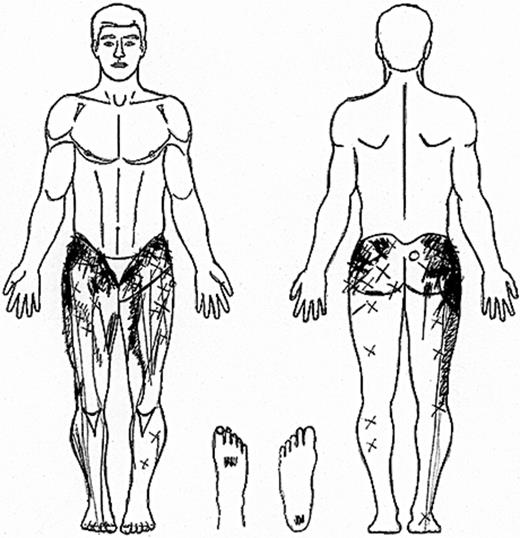
The researchers had 51 patients (28 female, 23 male) for their study. These patients had hip pathology as evidenced by x-ray or MRI. Pre injection, these patients marked out on a body chart where their pain was (see picture below).

Pain referred from the hip 
The patients were then given a fluoroscopically guided intra-articular (FGIA) injection. There was only local anesthesia and no oral or intravenous sedation for the patients to avoid confounding of results. Only when their pain was reduced by 90% before the FGIA injection was deemed effective. 

Long needle to reach the hip
The researchers found that referred pain to the buttock was the most common (71%). Traditionally accepted referral to the thigh (57%) and groin (55%) were less common. 22% of patients had referred pain to the legs (below the knee).

The referred pain areas in this study (from the hip joint) were similar to previously reported pain patterns observed from the lumbar spine and sacro iliac joints.

This is a very useful study for knowing referral patterns of the hip joint. When a patient complains of pain in their buttocks, groin, thigh or even in the foot, one must not rule out the hip. It is important to note that there is no lumbar spine referral in this study.

Note to self (and other physiotherapists reading this), not all radiating leg pain is 'sciatica'.

Reference

Lesher JM, Dreyfuss P, Hager N et al (2008). Hip Joint Pain Referral Patterns: A Descriptive Study. Pain Med. 9(1): 22-25. DOI: 10.1111/j.1526-4637.2006.00153.x

Friday, May 22, 2020

Physical Activities Does Not Wear Out Our Spine

I've written more than five other posts on our back / spines recently, well here's another article to align with our Instagram and Facebook posts this week. You can watch the three videos here.

We've all been sitting a whole lot more, myself included during the circuit breaker. Here's what research is suggesting, that it is actually physical inactivity and not physical labor that is associated with degenerative (or wear and tear) changes in the thoracic and lumbar spine.

The researchers studied 385 subjects on the relationship between short and long term physical inactivity and degenerative changes of the thoracic and lumbar spine over a period of 14 years. They grouped the subjects into those who did no physical activity, or did so irregularly for 1 hour a week, regularly for 1 hour a week, or regularly for more than 2 hours a week.

In addition, physical labor, walking and cycling were investigated additionally.

Correlations between physical inactivity and thoracic and lumbar disc degeneration were analyzed after accounting for sex, age, Body Mass Index, hypertension, diabetes and back pain.

Subjects with disc degeneration in the thoracic and lumbar spine were more common in those with no physical activity, irregular activity < 1 hour compared than those with regularly activity > 1 hour or more a week.

You'll be happy to know that there was no obvious significant association statistically for subjects who did physical labor, walking or cycling with disc degeneration.

The researchers concluded that physical inactivity over the 14 years they studied demonstrated a strong correlation with disc degeneration of the thoracic and lumbar spine.

Just like we wrote previously that running more miles does not wear out your knees, being active, doing physical labor will not wear out your spine too.

Keep moving.


Reference

Maurer E, Klinger C et al (2020). Long-term Effect Of Physical Inactivity On Thoracic And Lumbar Disc Degeneration- An MRI-based Analysis of 385 Individuals From The General Population. Spine DOI: 10.1016/j.spinee.2020.04.016


My boys wanted the same picture as me, so here's my older boy.


And here's my younger boy below.


Monday, February 3, 2020

What You Need To Know About Slipped Discs


We're still writing about our backs. This week I'm writing about 'slipped discs' in the spine. This is a topic that I get questions from most often among my friends and patients alike.

Patients have always been told that the intervertebral disc (IVD) causes referred pain down the back/ side of your legs. They often think that discs are very fragile and get easily injured.

Almost every single one of my patients are fearful of having a 'slipped intervertebral disc' whenever they have low back pain (LBP). The discs have a really bad reputation for causing significant pain and disability in many people.

Are these common beliefs accurate? Let's go through the anatomy of the spine and the IVD.


The IVD consists of a very tough outer layer called the annulus fibrosis (AF). It is made of of several layers of fibrocartilage consisting of Type I and II collagen fibers. The AF protects the soft, gel-like substance in the middle known as the nucleus pulposus (NP). The NP helps distribute pressure evenly across the IVD and prevent excessive forces on the spine.


See how thick the AF is from the picture above? Here's something else you need to know. There is a cartilaginous endplate between the AF and the vertebra (the spine). The endplates hold the IVD in place. It allows load to be spread evenly and to provide attachment to the IVD. This creates a super strong connection to the AF making it impossible for the IVD to 'slip' out of position.

How strong are our IVD's? In a published study on thoracic discs in the young (28 years old plus minus 8 years) , it took about 740 pounds of force to compress the disc height 1 mm. For the older subjects (70 years young plus minus 7 years), it took almost 460 pounds of force. Note that these are on cadavers with the muscles and bones cut away (Stemper et al, 2010).


The endplates also allow for hydration of the disc to take place (see picture above).

Just like your ACL is often injured by shearing forces, it is also shearing forces that is most likely to hurt your discs. Twisting, rotating your back while lifting a heavy load is definitely not recommended.

What happens after your disc is injured? Find out more next week as I write more on that topic.


Reference

Stemper BD, Board D et al (2010). Biomechanical Properties Of Human Thoracic Spine Disc Segments. J Craniovert Junct Sp. 1(1): 18-22. DOI: 10.4103/09774-8237.65477

Monday, January 27, 2020

Transverse Abdominis and Low Back Pain

I remember being a second year physiotherapy student in the early 90's when we had a special tutorial on how to assess and strengthen the Transverse Abdominis (TA). While in crook lying, you move your leg (bent knee fall out), leg straightening etc and had to maintain the TA contraction.

This was just before a young Paul Hodges and his supervisor Carolyn Richardson (both very famous physiotherapists) published his article on the TA. His research compared the timing of TA and Multifidus in people with low back pain (LBP) for 18 months or more against healthy subjects with no LBP.

Using fine wire and surface EMG, the participants had to move their leg or shoulder and their timing of TA and Multifidus muscles measured. Those with LBP had delayed activation of their TA/ Multifidus.
Hodges suggested that the TA was the likely to be the main cause of LBP as it is the most important and deepest muscle in the abdomen. It looks and works like a corset to stabilise the back.

This article made the allied healthcare/ fitness industry conclude that the TA was a trunk stabilising muscle that was very important to strengthen for those with LBP. Patients who had LBP had weaknesses in their TA which led to instability in the spine.

Because of Hodges' research, many other research on LBP went in that direction. However, out of the seven systematic reviews found, six of them now showed no increase in benefit in such TA based exercises over general exercises. Further research is probably not going to significantly alter this.

As I wrote last week, this early work by Hodges, Richardson and Gwen Jull was quickly adopted by the fitness industry (though Joseph Pilates never intended this). The Allied Health/ Physiotherapy community started assessing the TA via palpation, ultrasound etc and coming up with various rehabilitation exercises for it as well. There are various continuing education courses teaching this approach still.

Although research says this may help, it is no better than general graded exercise and we know that LBP is also associated with obesity and mental health (Maher et al, 2016).

There is even research advising those who have been trained to brace/ hollow their 'core' should be discouraged from doing this as this may create abnormal movement patterns creating more stress on the spine (Lederman et al, 2010).
Brace or hollow your core?
So rather than getting our patients to feel for a deep muscle (like the TA and Multifidus) in a low load movement, it would make more sense for us to get patients to do an exercise/ activity they enjoy that gives them similar benefits.


References

Hodges PA and Richardson CA (1998). Delayed Postural Contraction Of Transversus Abdominis In Low Back Pain Associated With Movement Of The Lower Limb. J Sp Disorders. 11(1): 46-56.

May S and Johnson R (2006). Stabilisation Exercises For Low Back Pain: A Systematic Review. Physiotherapy. 94(3): 179-189. DOI: 10.1016/j.physio.2007.08.010

Maher CG, Underwood M et al (2016). Non-specific Low Back Pain. The Lancet. 389(10070): 736-747. DOI: 10.1016/S0140-6736(16)30970-9

Lederman E (2010). The Myth Of Core Stability. J of Bodywork, Mvt Ther. 14(1); 84-98. DOI: 10.1016/jbmt.2009.08.001

Saragitto B, Maher CG et al (2016). Motor Control Exercise For Nonspecific Low Back Pain. A Cochrane Review. Spine 41(16): 1288-1295. DOI: 10.1097/BRS.0000000000001645

Friday, September 8, 2017

Lumbar Spondylolisthesis But No Real Deficits So Far


Have a look at the MRI and you can see a very obvious spondylolisthesis in my 38 year old patient's lumbar spine at the L5-S1 area. The last time I saw one so obvious was when I was working at the Singapore Sports Institute in a young gymnast in 2001.

Spondylolisthelis is a condition in which one vertebra slides forward over the vertebra below it. In simple terms, it means that there is a forward slip of one vertebra relative to another.

From what my patient says, there wasn't any traumatic incidents. He complains of lumbar area aches in the past year.  Often there is discomfort over his L buttock and tightness down his L outer thigh at the end of the day. He is still able to exercise including running and playing football. Other than that he hardly has any other symptoms.

His surgeon has suggested surgical intervention to stabilize his spine at the end of next month. Looks like lots of rehab after surgery.


Monday, May 29, 2017

Steroid Injection Not Better Than Placebo For Low Back Pain

Picture by Army Medicine from Flickr
It seems like another of my patients has been asked if he wanted a cortisone (or corticosteroid) injection for his low back pain after I wrote about my shoulder pain patient last week who was asked by another doctor to get a steroid injection.

Evidence that they (cortisone or corticosteroid injections) work no better than placebo is mounting though.

The article referenced below is a systematic review. A systematic review is a computer aided search for all randomized and clinical controlled trials, meaning it's top of the line in terms of quality.

Researchers pooled data together from 30 placebo controlled studies of epidural steroid injections for radiculopathy (back pain that radiates to legs) and eight studies for spinal stenosis (back/ neck pain caused by narrowing of the spinal canal).

For radiculopathy, the steroid injections provided only short term relief. Over time, they were not more effective compared to placebo injections. They also did not reduce the need for surgery later for patients with more severe conditions.

The pooled data was similar for spinal stenosis.There was only some moderate temporary pain relief after the corticosteroid injections. There were no differences between steroid injections and placebo injections in pain intensity or functional ability lasting six weeks or longer after the injections.

The authors did note that some studies did not have many subjects while others had shortcomings their methodology.

The researcher in charge also suggested that there was probably some financial motivation to carry on using steroid injections despite the lack of evidence for its effectiveness. There is also a worry among professional societies (for doctors) due to implications for insurance coverage.

Other mentioned less selfish reasons is that doctors see their patients in horrible pain and concede that they do get some pain relief after the injections.

Please come to our clinics if you need help with your low back pain. We definitely do not give steroid injections.


Reference

Chou R, Friedly J et al (2015). Epidural Corticosteroid Injections For Radiculopathy And Spinal Stenosis: A Systematic Review And Meta-analysis. Ann Intern Med. 163(5): 373-381. DOI: 10.7326/M15-0934.